EndoTODAY 내시경 교실
Beginner | ESA | Schedule | OPD
Seminars | Atlas | Recent | Links
![]() [Thursday Endoscopy Conference 20161222]
[Thursday Endoscopy Conference 20161222]
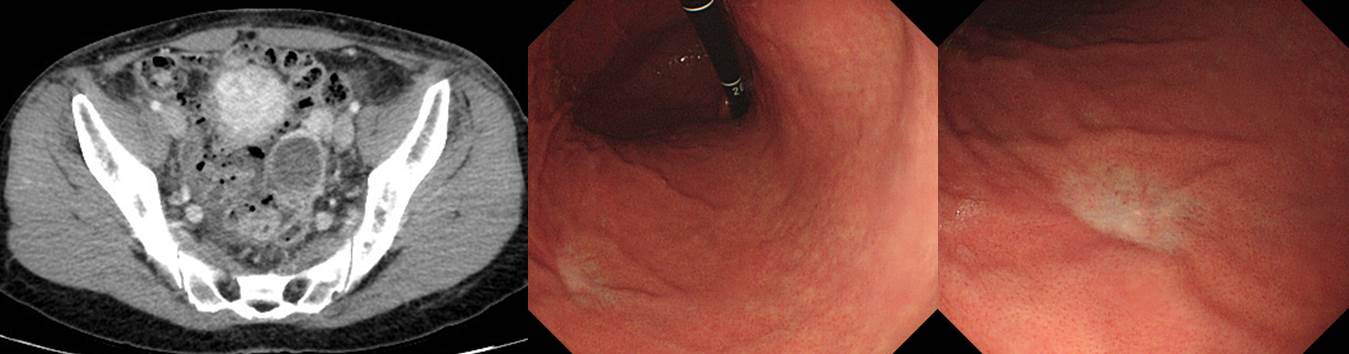
![]() 1. Small signet ring cell carcinoma with peritoneal seeding and ovarian metastasis
1. Small signet ring cell carcinoma with peritoneal seeding and ovarian metastasis
Left ovarian mass로 산부인과 수술 후 signet ring cell carcinoma로 나와 primary site workup 위하여 의뢰되었음. 위내시경에서 위체하부 전벽의 small pale depressed lesion (blurred edge, flat margin, no converging fold)이 발견되었고 조직검사에서 signet ring cell carcinoma가 나옴. 작은 위암이 전이된 상태였던 것으로 잠정 진단함.
오래 전에도 비슷한 증례가 있었습니다. 사실 EGC 아닌가 생각했는데 AGC였고 multiple lymph node 전이와 bilateral ovary metastasis까지 있었습니다.
(2012년, 50세 여성)
Stomach, radical subtotal gastrectomy: Advanced gastric carcinoma
1. Location : middle third, Center at angle and posterior wall
2. Gross type : Borrmann type 3
3. Histologic type : tubular adenocarcinoma, poorly (solid) differentiated >> mucinous adenocarcinoma (mucinous carcinoma portion: 20%)
4. Histologic type by Lauren : mixed
5. Size : 3.3x3.0 cm
6. Depth of invasion : penetrates subserosal connective tissue (pT3)
7. Resection margin: free from carcinoma, safety margin: proximal 3.3 cm, distal 3.8 cm
8. Lymph node metastasis : metastasis to 6 out of 32 regional lymph nodes (pN2) (perinodal extension: present) (6/32: "1", 0/2; "3", 2/2; "4", 2/9; "5", 0/0; "6", 2/5; "7", 0/4; "9", 0/6; "8a", 0/3; "11p", 0/1; "12a", 0/0; "4sb", 0/0)
9. Lymphatic invasion : present
10. Venous invasion : not identified
11. Perineural invasion : not identified
12. AJCC stage by 8th edition: pT3 N2Ovary and salpinx, bilateral salpingo-oophorectomy : METASTATIC CARCINOMA, clinically from stomach
Location: Bilateral ovaries
Greatest dimension: 5.5 cm (left)
Lymphovascular invasion: PRESENT (focal)
Salpingeal extension: Absent (lymphovascular invasion only; left salpinx)
약간 다르지만 아래 동영상은 보만 4형 진행성 위암 난소 전이 증례들에 대한 상세한 설명입니다.

* 참고: 난소에 전이를 일으킨 위암의 임상적, 병리학적 고찰 대한소화기학회지 1998;32:725-732
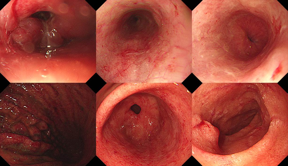
![]() 2. Corrosive esophagogastroduodenitis
2. Corrosive esophagogastroduodenitis
2시에 락스를 마신 후 8시에 응급실 방문. 9시 30분에 내시경 시행
부식성 식도염에서 꼭 내시경 검사를 해야 하는가에 대한 짧은 토론이 있었습니다. 일찍 내시경 검사를 하는 것은 손상의 정도를 평가하고 예후를 예측하는데 도움이 될 것이라는 의견이 많았습니다. 저도 동의하였습니다. 다만, 조금이라고 애매하면 검사하지 않거나 검사를 중간에 중단해도 무방할 것 같다고 생각합니다. 무리할 필요는 없을 것 같습니다.
* 참고: EndoTODAY 부식성 식도염
![]() 3. Upper esophageal diverticulum
3. Upper esophageal diverticulum
항상 쉬운 것은 아닙니다. 아래 증례를 보십시오.
목의 통증으로 내시경 검사를 시행하여 상부식도의 erythematous swelling, stenosis 소견만 있었고 조직검사에는 염증 소견만 나왔고 neck CT에서 암 의심으로 의뢰되신 분입니다.
첫 내시경. 가는 내시경으로 바꾸어 통과할 수 있었음.
의뢰 후 내시경 재검에서 UES 직하방에 blind end를 이루는 pouch가 있었고, 이 pouch의 proximal 측면에 작은 opening이 관찰됨. 이 구멍을 통하여 식도 true lumen으로 들어갈 수 있었음.
상부식도 게실에 음식이 차 있고 true lumen으로 들어가는 길을 찾기 어려웠기 때문에 상부식도의 악성질환으로 오인되었던 것 같습니다. 고령 환자의 상부식도 질환에서는 반드시 diverticulum을 고려해야 합니다.
최근에는 내시경 치료가 활발히 이루어지고 있습니다.
참고: EndoTODAY Zenker's diverticulum
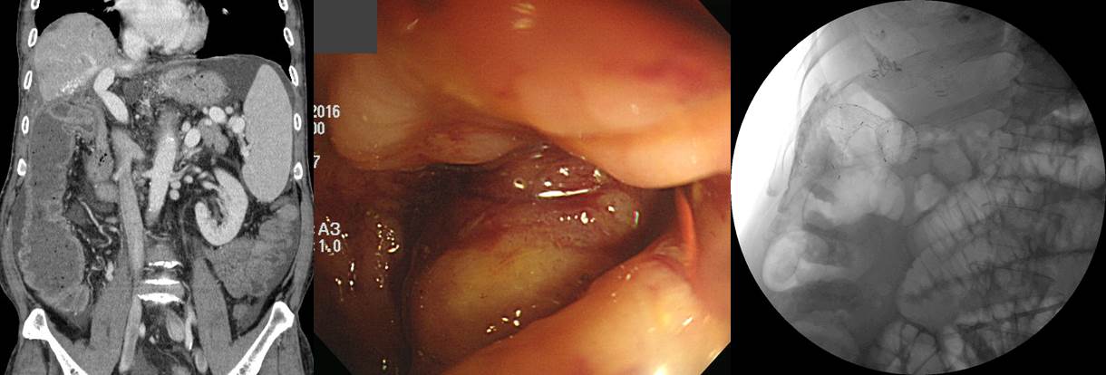
![]() 4. HCC with colonic invasion
4. HCC with colonic invasion
HCC로 오래 치료받던 환자가 복통, 변비, 복부 팽만감으로 내원하여 대장내시경을 하였을 때 colonic obstruction 소견으로 stent를 하였고 일시적인 증상의 완화를 보였습니다.
* 참고: EndoTODAY GI involvement of HCC
![]() [References]
[References]
1) SMC Endoscopy Unit 삼성서울병원 내시경실
2) SMC Monday GI conference 삼성서울병원 일원내시경교실 월요점심소화기집담회
3) SMC Thursday endoscopy conference 삼성서울병원 일원내시경교실 목요점심내시경집담회
© 일원내시경교실 바른내시경연구소 이준행. EndoTODAY Endoscopy Learning Center. Lee Jun Haeng.