EndoTODAY 내시경 교실
Beginner | ESA | Schedule | OPD
Seminars | Atlas | Recent | Links
![]() [Thursday Endoscopy Conference 20170112]
[Thursday Endoscopy Conference 20170112]
![]() 1. EGC-like AGC
1. EGC-like AGC
Advanced gastric carcinoma
1. Location : upper third, Center at body and posterior wall
2. Gross type : Borrmann type 3
3. Histologic type : tubular adenocarcinoma, poorly (poorly cohesive) differentiated
4. Histologic type by Lauren : diffuse
5. Size : 3.7x2.8 cm
6. Depth of invasion : penetrates subserosal connective tissue (pT3)
7. Resection margin: free from carcinoma, safety margin: proximal 2.7 cm, distal 11.7 cm
8. Lymph node metastasis : metastasis to 3 out of 38 regional lymph nodes (pN2)
9. Lymphatic invasion : present(+++)
10. Venous invasion : not identified
11. Perineural invasion : not identified
12. Peritoneal cytology : negative
13. AJCC stage by 7th edition: pT3 N2
* 참고: EndoTODAY 심달도
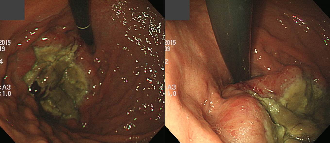
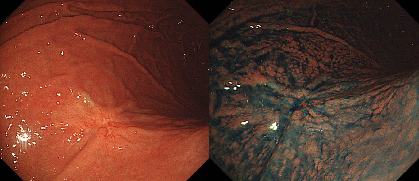
![]() 2. Localized colon amyloidosis
2. Localized colon amyloidosis
* 참고: EndoTODAY 대장 아밀로이드증, EndoTODAY 위 아밀로이드증
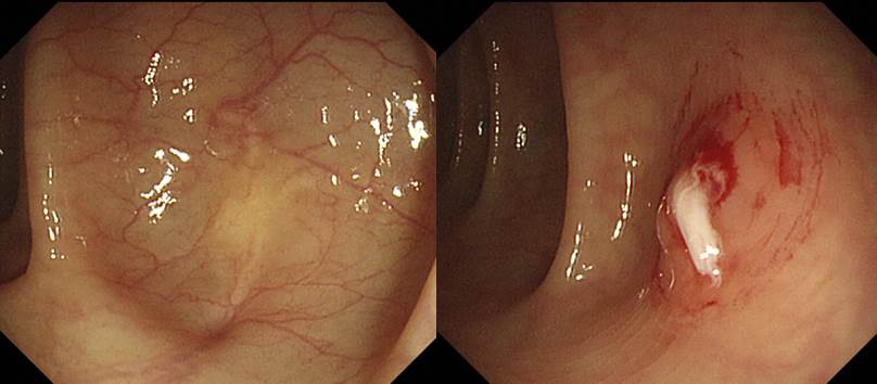
![]() 3. Gastric metastasis from malignant melanoma
3. Gastric metastasis from malignant melanoma
두통을 주소로 검사하여 lung cancer with brain metastasis로 의뢰된 70대 남성입니다. Lung biopsy (gun)은 "poorly differentiated tumor with dyscohesive anaplastic cells. The possibility of malignant melanoma is suspected."였습니다.
위내시경 조직검사는 아래와 같았습니다.
Malignant melanona.
S-100: diffusely positive in tumor cells
Melanoma Ag (HMB45): diffusely positive in tumor cells
c-kit gene, BRAF gene: no mutation
이 환자는 skin lesion은 없었습니다. 따라서 찾지 못하는 skin melanoma의 lung, brain, stomach metastasis일 수도 있고, primary lung melanoma의 multiple metastasis일 수도 있을 것 같습니다. Stage가 너무 높아 치료 계획이 달라질 것은 없지만... 내시경의사로서 특이한 점은 melanoma stomach metastasis의 색조가 검지 않았다는 점입니다.
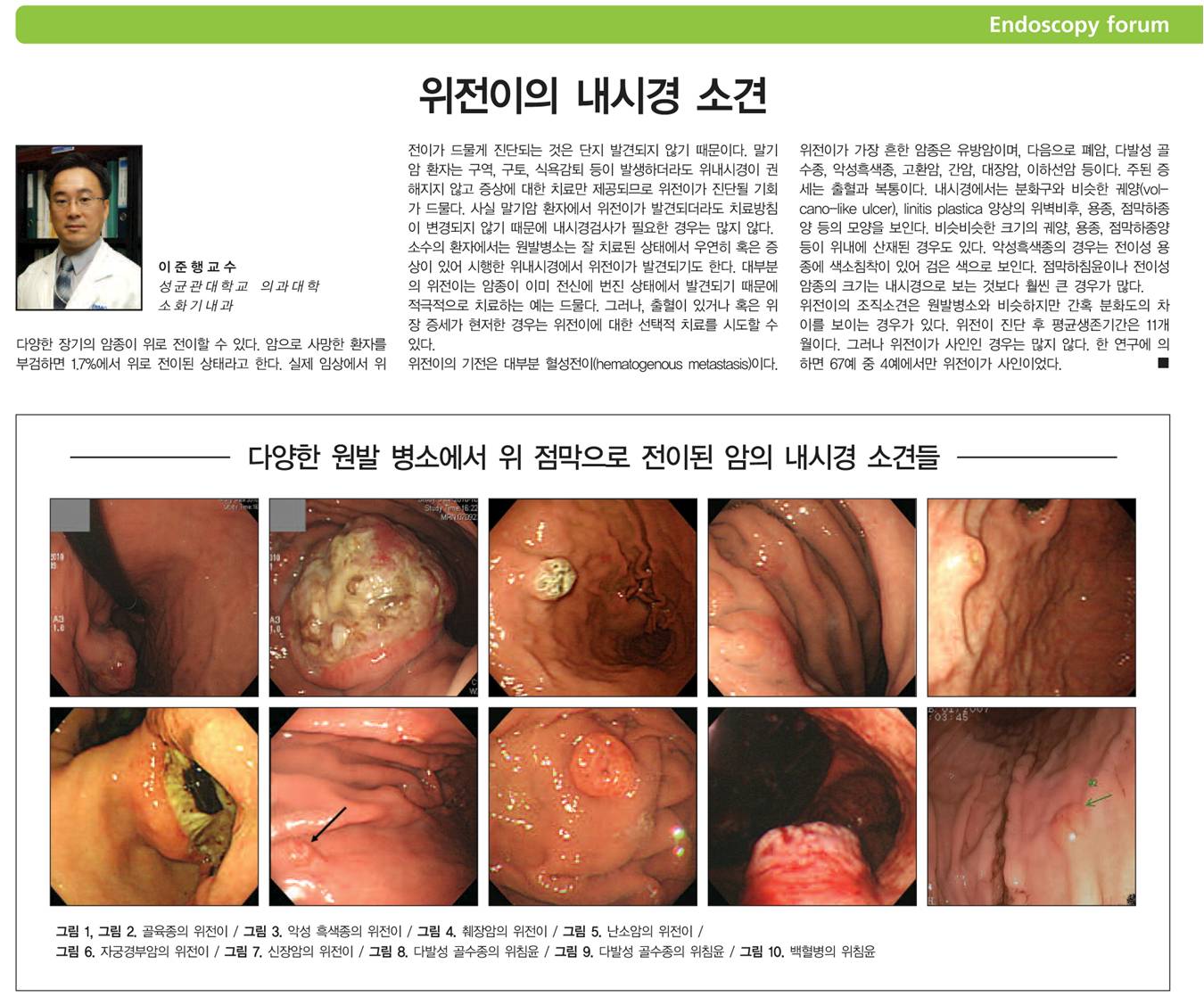
다양한 악성질환이 위로 전이할 수 있습니다. 수년 전 Elsevier에서 나오는 GI & Hepatology News (Korean Edition)에 기고한 내용입니다.
흑색종 위전이에 대하여 과거 소개한 내용을 옮깁니다. 우리 나라에서는 서구에 비하여 흑색종이 드문 편이지만, 비교적 진행된 상태로 발견되는 예가 많아서 예후는 불량합니다. 흑색종은 소화관으로 잘 전이되는 대표적인 악성질환으로, 흑색종으로 사망한 환자의 60%가 부검에서 소화관 전이가 발견됩니다. 가장 전이 빈도가 높은 곳은 소장이지만 대장, 위, 식도 등에도 전이가 흔합니다. 위로의 전이는 약 10-26% 정도이며 다발성 점막하 종양으로 관찰됩니다. 특히 점막하 종양 상단의 depression과 black discoloration이 특징입니다. 간혹 amelanotic melanoma도 있으므로 (이 경우는 black이 아니다) 주의할 필요가 있습니다. 광범위하게 전이된 흑색종의 10% 정도에서는 원발부위를 찾을 수 없습니다 (대한소화기내시경학회지 2004;28:71-75).
두통과 blurred vision으로 내원한 환자입니다. 뇌 MRI에서 “Malignant bone mass such as skull base metastasis with hypoglossal canal involvement and denervation injury of tongue, right”의 소견이었고 PET에서 multiple metastasis가 관찰되었습니다. 위내시경에서 위체부와 전정부에서 골고루 매우 많은 검은색 점들이 관찰되었고 조직검사에서 malignant melanoma, HMB45 (+), S-100 (Focal +), cytokeratin (-)로 확인되었습니다. 등에서 점이 발견되었고 조직검사에서 malignant melanoma였습니다. Multiple metastasis가 의심되었던 환자에서 위내시경을 통하여 melanoma가 확인된 후 전신에 대한 자세한 신체검진을 통하여 원발병소를 발견한 경우입니다. Melanoma의 위전이는 주로 SMT-like한 elevated lesion이 dark pigmentation된 형태로 보입니다. 오늘의 증례는 flat dark spot들로 관찰된 드문 예입니다.
얼굴 피부 흑색종 위전이
흑색종 십이지장전이, 뇌전이
흑색종 위전이로부터의 출혈

* 참고: EndoTODAY 위전이
![]() 4. Gastic lymphoma (DLBCL)
4. Gastic lymphoma (DLBCL)
아래 슬라이드를 보시기 바랍니다.
PPT PDF 4.6 M
* 참고: EndoTODAY 위장관 림프종
![]() [References]
[References]
1) SMC Endoscopy Unit 삼성서울병원 내시경실
2) SMC Monday GI conference 삼성서울병원 일원내시경교실 월요점심소화기집담회
3) SMC Thursday endoscopy conference 삼성서울병원 일원내시경교실 목요점심내시경집담회
© 일원내시경교실 바른내시경연구소 이준행. EndoTODAY Endoscopy Learning Center. Lee Jun Haeng.