EndoTODAY 내시경 교실
Beginner | ESA | Schedule | OPD
Seminars | Atlas | Recent | Links
![]() [Thursday Endoscopy Conference 20170126]
[Thursday Endoscopy Conference 20170126]
![]() 1. Radiation-related esophagitis
1. Radiation-related esophagitis
NSCCL (right lower lobe, squamous cell carcinoma, cT1bN3)로 CCRT 받은 환자의 odynophagia. IC 30cm 부위의 궤양. 조직검사: inflamed granulation tissue with activated endothelial cells, consistent with radiation-induced esophagitis
* 참고: EndoTODAY 방사선 관련 위장관 손상
![]() 2. Multiple synchronous cancer missed at the initial endoscopy
2. Multiple synchronous cancer missed at the initial endoscopy
위체중부 위암으로 의뢰된 환자를 subtotal gastrectomy로 치료하기 위하여 pre-op clipping 내시경을 하는 과정에서 위체상부에서 fundus로 넘어가는 부위의 또 다른 위암이 발견되었습니다. 어쩔 수 없이 total gastrectomy가 시행되었습니다. 처음 발견되었던 암보다 두번째 발견된 암이 더 크고 깊었습니다. 위암은 항상 다발성일 수 있습니다.
左: 처음 발견된 암, 中: 추가로 발견된 암, 右: clipping
Multiple gastric carcinomas (x2)
I. Early gastric carcinoma (mass 1) - 이것이 처음 발견된 암이었습니다.
1. Location : upper third, Center at body and posterior wall
2. Gross type : EGC type IIc
3. Histologic type : signet-ring cell carcinoma
4. Histologic type by Lauren : mixed
5. Size : 2.2x1.4 cm
6. Depth of invasion : invades mucosa (muscularis mucosae) (pT1a)II. Early gastric carcinoma (mass 2) - 이것이 clipping 과정에서 추가로 발견된 암이었습니다.
1. Location : middle third, Center at body and lesser curvature
2. Gross type : EGC type IIc
3. Histologic type : tubular adenocarcinoma, poorly (poorly cohesive) differentiated
4. Histologic type by Lauren : mixed
5. Size : 3x2.8 cm
6. Depth of invasion : invades submucosa (sm3) (pT1b)III.
1. Lymph node metastasis : no metastasis in 49 regional lymph nodes (pN0), (0/49: "3,5", 0/19; "4,6", 0/4; "2", 0/5; "5", 0/0; "6", 0/0; "7", 0/4; "9", 0/2; "8a", 0/5; "11p", 0/2; "12a", 0/5; "4sb", 0/1; "1", 0/2; "10", 0/0)
2. Lymphatic invasion : present
3. Venous invasion : not identified
4. Perineural invasion : not identified
5. Separate lesions : leiomyomas (x2) (size: 0.4x0.2 cm and 0.3x0.2 cm)
6. Resection margin : free from carcinoma, safety margin : proximal 5cm, distal 5.5cm
7. AJCC stage by 7th edition: pT1b N0
![]() 3. TB colitis
3. TB colitis
대장암 수술 후 추적 내시경 입니다. Ascending colon에서 우연히 결핵성 대장염이 발견되었습니다. 조직검사에서는 focal active colitis with focal crypt abscess 정도의 소견이었지만 tissus nested PCR 에서는 M. tuberculosis가 나왔습니다. 무증상 결핵성 대장염도 반드시 치료해야 하는가 논란이 가능하지만, 이런 경우 저는 보통 치료를 권하고 있습니다.
[Intestinal Tb 가 의심될 경우 조직검사 처방]
1) BL1A112A. colon 생검 1~3개 → 포르말린 통
2) BL4112. AFB stain and culture 처방 (검체 others(specify)) → saline 통
3) BG510101. Mycobacterium tuverculosis,nested PCR (검체 P17 colon) → 생검조직만
조직검사하기 전 미리 간호사에게 정보를 주어야 포르말린통에 검체가 모두 담기는 것을 막을 수 있습니다. 꼭 미리 소통하세요.
* 참고: EndoTODAY 결핵성 대장염
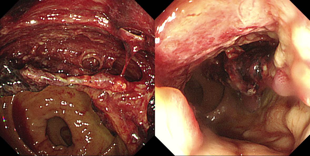
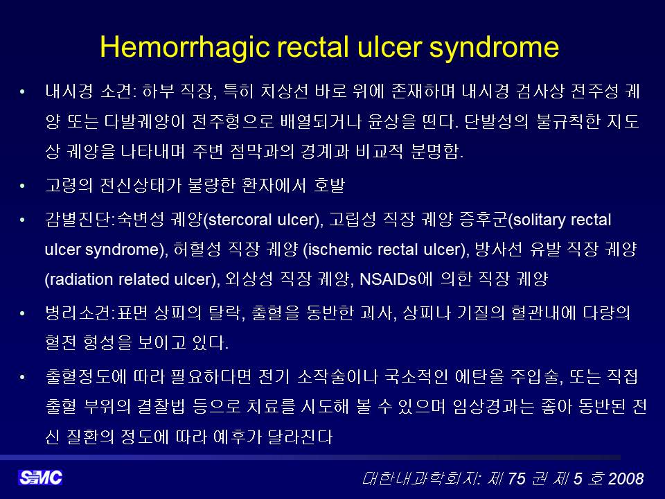
![]() 4. Hemorrhagic rectal ulcer syndrome
4. Hemorrhagic rectal ulcer syndrome
술을 자주 드시는 당뇨환자께서 음주 후 선홍색 혈변으로 내원하셨습니다.
![]() [References]
[References]
1) SMC Endoscopy Unit 삼성서울병원 내시경실
2) SMC Monday GI conference 삼성서울병원 일원내시경교실 월요점심소화기집담회
3) SMC Thursday endoscopy conference 삼성서울병원 일원내시경교실 목요점심내시경집담회
© 일원내시경교실 바른내시경연구소 이준행. EndoTODAY Endoscopy Learning Center. Lee Jun Haeng.