EndoTODAY 내시경 교실
Beginner | ESA | Schedule | OPD
Seminars | Atlas | Recent | Links
![]() [Thursday Endoscopy Conference 20170427]
[Thursday Endoscopy Conference 20170427]
![]() 1. Double primary cancer (?)
1. Double primary cancer (?)
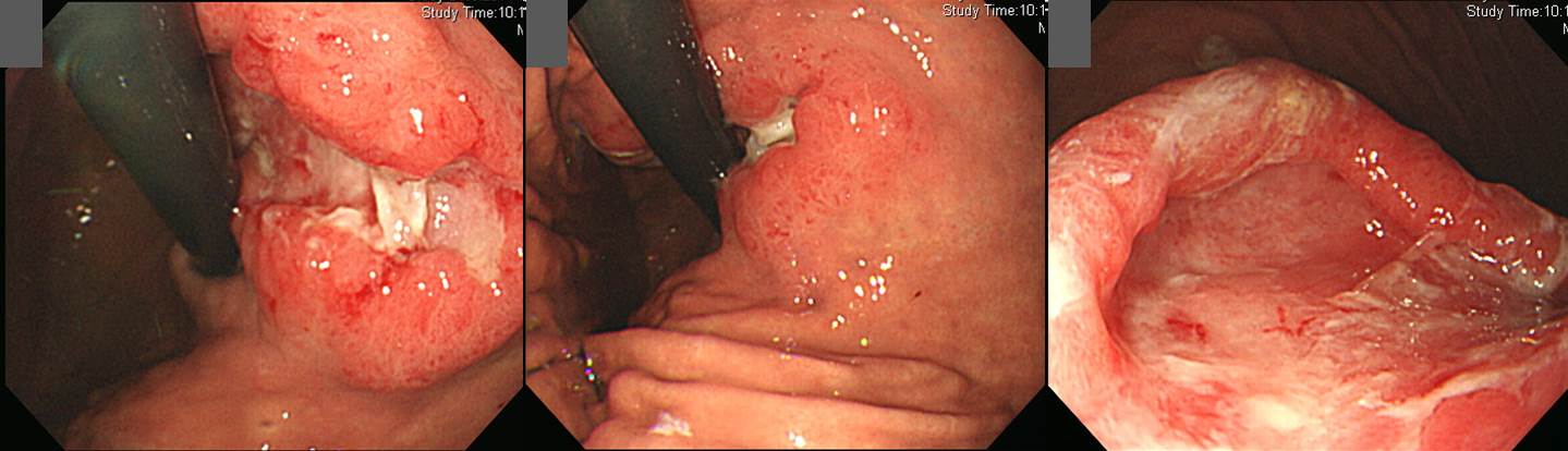
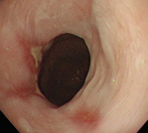
폐암 (adenocarcinoma), pleural seeding으로 clinical trial을 위하여 의뢰되었던 환자입니다. 초기 평가 과정에서 위의 종양이 발견되었습니다.
위 조직검사는 tubular adenocarcinoma (poorly differentiated)로 확인되었습니다. 원발성 위암과 폐암의 위전이의 구분이 쉽지 않은 증례입니다. 후향적으로 과거 사진을 검토해 보았을 때 2년 전 처음 폐암 진단될 당시의 내시경 사진에서도 같은 부위에 점막병소가 있는 것으로 추정되었습니다.
폐암의 위전이와 원발성 위암의 구분이 쉽지 않겠으나, 일단 원발성 위암의 가능성이 더 높을 것으로 추정하였습니다. 정답은 아무도 모르지만...
* 참조: EndoTODAY 위전이
![]() 2. AGC Borrmann type IV with pseudopyloric ring
2. AGC Borrmann type IV with pseudopyloric ring

Stomach, total gastrectomy:
Advanced gastric carcinoma
1. Location : entire stomach, Center at body and anterior wall
2. Gross type : Borrmann type 4
3. Histologic type : tubular adenocarcinoma, poorly differentiated
4. Histologic type by Lauren : diffuse
5. Size : 15x13 cm
6. Depth of invasion : invades serosa (pT4a)
7. Resection margin: free from carcinoma, safety margin: proximal 1.5 cm, distal 2 cm
8. Lymph node metastasis : metastasis to 5 out of 50 regional lymph nodes (pN2), (perinodal extension: absent) (5/50: "LN3,5", 0/2; "LN4,6", 2/5; "1", 0/3; "2", 0/4; "4ss", 1/7; "5", 0/1; "6", 0/11; "8a", 0/4; "7", 0/1; "9", 1/2; "11p", 0/1; "12a", 0/3; perigastric, 1/6)
9. Lymphatic invasion : present (++)
10. Venous invasion : not identified
11. Perineural invasion : present
12. Peritoneal cytology : atypical cells
13. AJCC stage by 7th edition: pT4a N2
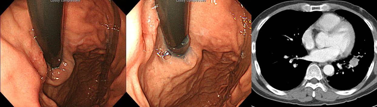
Fundus와 high body는 비교적 extensibility를 유지하고 있으나 위체하부와 근위 전정부는 잘 펴지지 않고 그 부위에 작은 궤양 병소가 관찰되었습니다. 조직검사에서 암이 확인되어 수술을 하였습니다. 비록 조직검사에서 암이 확인되지 않았더라도 적극적인 수술이 필요한 경우라고 판단됩니다.
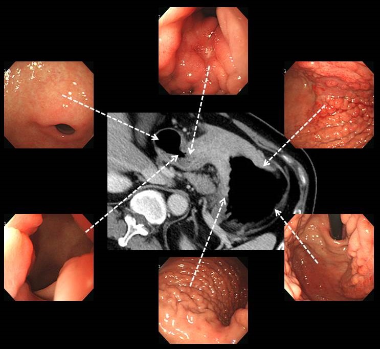
집담회 과정 중 내시경 사진만 보고 위암의 3차원적 구조를 잘 이해하지 못한 분들이 계셔서 CT gastrography 영상을 이용하여 그림을 그려 보았습니다. 위 fundus는 비교적 save 되었고, 위체상부부터 근위전정부까지 diffuse하게 infiltration하는 양상입니다. 위체중부부터는 현저히 좁아져 있고 근위전정부에서는 거의 막히기 직전까지 간 상태입니다. 원위 전정부는 save 되어 있습니다. 따라서 좁아진 부위 끝이 마치 pseudo-pyloric ring 비슷한 모양을 이루고 있습니다.
* 참고 1: EndoTODAY 보만 4형 진행성 위암
* 참고 2: Borrmann type IV AGC. Clin Endosc 2016
![]() 3. Reflux esophagitis with ulcer and stricture, Sliding hiatal hernia
3. Reflux esophagitis with ulcer and stricture, Sliding hiatal hernia
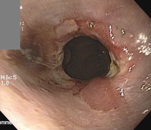
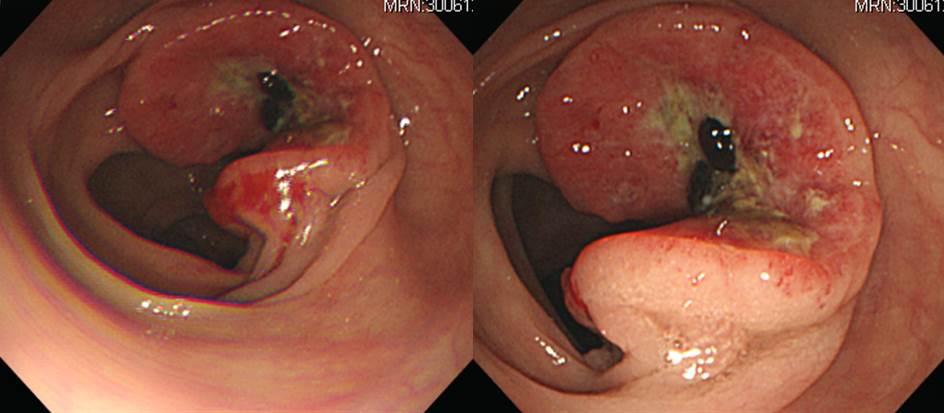
사진 한장입니다. 진단은?
집담회에서는 의견이 갈려서 GERD라고 한 분도 있고 암의 가능성이 있는 의견을 피력한 분도 있었습니다. 진료를 담당하였던 민양원 교수님은 PPI를 투약하고 한 달 후 내시경 재검을 받도록 하였습니다. 한 달 후 사진은 아래와 같습니다.
PPI 한 달 투여 후 사진
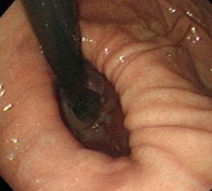
Hiatal hernia가 있고 mucosal ring 높이에서 약간의 stricture와 함께 marginal erosion과 ulceration이 있었던 경우로 PPI 투약후 곧 호전되었습니다. 그렇다면 교수님은 어떻게 암이 아니라는 것을 그다지도 확신을 가지고 진료를 하셨던 것일까요? 답은 아래에 있습니다. 3년 전 내시경에서도 거의 비슷한 소견이 있었던 것입니다. 단지 stricture 소견이 전혀 없었을 뿐입니다. 8시 방향의 gastric fold의 proximal end가 약간 부풀어 올라서 용종처럼 보이는 부분이 있습니다. 100% benign, sentinel polyp이라고 할 수 있는 것입니다.
3년 전 사진
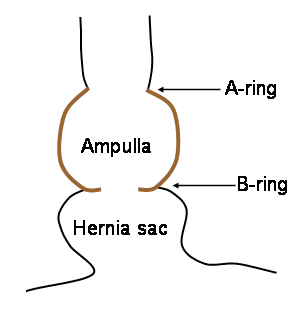
거의 비슷한 증례입니다. 아래는 hiatal hernia가 있으면서 squamocolumnar junction 부위 (B ring, mucosal ring)가 좁아지면서 scar를 형성하고 있는 증례입니다. Erosion들이 longitudinal한 방향보다는 좁아진 축을 따라서 circumferential하게 형성되어 있고 일부는 triangle shape입니다. Marginal erosion이라고 부르기도 합니다.
![]() 4. Colon cancer
4. Colon cancer
Transverse colon, left hemicolectomy:
Adenocarcinoma, poorly differentiated
1. Location: transverse colon
2. Gross type: ulcerofungating
3. Size: 4x3 cm
4. Depth of invasion: penetrates visceral peritoneum(pT4a)
5. Resection margin: free from carcinoma, safety margin: nearest, 4 cm ; opposite, 11 cm
6. Regional lymph node metastasis : metastasis to 6 out of 14 regional lymph nodes(pN2) (6/14: pericolic, 6/14)
7. Lymphatic invasion: present
8. Venous invasion: not identified
9. Perineural invasion: not identified
10. Tumor budding : negative
11. Pathologic staging: T4a N2 Mx
![]() 5. Diffuse large B cell lymphoma
5. Diffuse large B cell lymphoma
7 주 간격으로 두 번 시행한 내시경에서 종양성 질환이 의심되는데 조직검사에서 암으로 나오지 않아 의뢰된 환자입니다.
첫 내시경. Biopsy: R/O Activated lymphocytes or lymphoma LGD
두번째 내시경. Biopsy: chronic gastritis
의뢰 후 내시경 소견도 두번째 내시경과 비슷하였고 조직검사를 열심히 했는데 이번에는 림프종으로 진단이 되었습니다.
Biopsy: diffuse large B cell lymphoma
Diffuse large B cell lymphoma는 Helicobacter gastritis와 연관된 예가 많습니다. 이 환자의 첫 내시경을 보면 위체상부와 fundus에 Helicobacter-associated gastritis에 의한 diffuse redness가 현저하고, 전정부에는 lymphofollicular gastritis가 관찰됩니다. 이 모든 것이 Helicobacter와의 관련성을 시사하는 소견입니다. 비단 adenocarcinoma 예방뿐만 아니라 여러 이유로 Helicobacter는 제균해 주는 것이 좋겠다는 것이 제 생각입니다.
/적/응/증/확/대/하/라/
* 참고: EndoTODAY Diffuse large B cell lymphoma
![]() [References]
[References]
1) SMC Endoscopy Unit 삼성서울병원 내시경실
2) SMC Monday GI conference 삼성서울병원 일원내시경교실 월요점심소화기집담회
3) SMC Thursday endoscopy conference 삼성서울병원 일원내시경교실 목요점심내시경집담회
© 일원내시경교실 바른내시경연구소 이준행. EndoTODAY Endoscopy Learning Center. Lee Jun Haeng.