 EndoTODAY 내시경 교실
Beginner | ESA | Schedule | OPD
Seminars | Atlas | Recent | Links
![]() [위암 196 - SM3 cancer]
[위암 196 - SM3 cancer]
001 | 101 | 201 | 301 | 401 | 501 | 601 | 701 | 801 | 901 | 1000
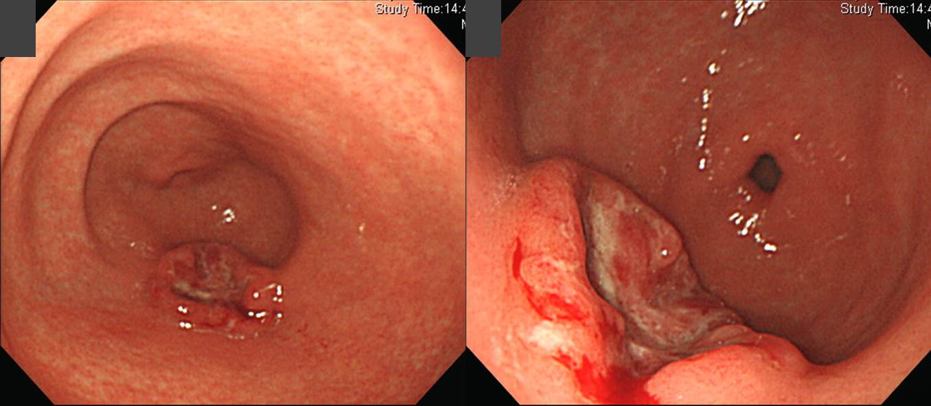
Early gastric carcinoma
1. Location : lower third, center at antrum and greater curvature
2. Gross type : EGC type IIc+IIa
3. Histologic type : lymphoepithelioma-like carcinoma (EBV-positive)
4. Histologic type by Lauren : intestinal
5. Size : 2.5x2x0.5 cm
6. Depth of invasion : extension to submucosa (sm3) (pT1b)
7. Resection margin: free from carcinoma, safety margin: proximal, 10.0 cm; distal, 1.8 cm
8. Lymph node metastasis : metastasis to 2 out of 36 regional lymph nodes (pN1)
9. Lymphatic invasion : not identified
10. Venous invasion : not identified
11. Perineural invasion : not identified
![]() 이 질환의 임상적 특징에 대해서는 최근 삼성서울병원 외과의 아래 논문을 참고하시기 바랍니다 (J Surg Res 2014)
이 질환의 임상적 특징에 대해서는 최근 삼성서울병원 외과의 아래 논문을 참고하시기 바랍니다 (J Surg Res 2014)
Lymphoepithelioma-like carcinoma: A distinct type of gastric cancer.
Lymphoepithelioma-like carcinoma (LELC) is a rare type of gastric carcinoma and has histologic features of intense lymphocytic infiltration. In this study, we attempted to analyze the clinicopathologic characteristics and survival outcome of patients with LELC compared with those with non-lymphoepithelioma-like carcinoma (NLELC).
METHODS: We studied 4282 patients who underwent gastrectomies to treat gastric cancer at the Department of Surgery of the Samsung Medical Center in Seoul, between January 2008 and December 2010. The clinicopathologic features and clinical outcomes of patients with LELC (n = 46) were compared with those with NLELC (n = 4236). In situ hybridization for Epstein-Barr virus (EBV) positivity was performed on the tissue of patients with LELC (n = 46) and NLELC (n = 1247).
RESULTS: The patients with LELC are male predominant and had more upper locations, more indeterminate Lauren classifications, lower T stages, less lymphatic invasion, and more positive EBV in situ hybridization compared with those of the NLELC group (80.4% versus 6.5%). Age, histologic type, Lauren type, the location of the tumor, the depth of the invasion, lymph node metastasis, and venous invasion were independent prognostic factors; however, the LELC type itself was not predictive of outcome. The 5-y survival rate of the LELC group (97.7%) was better than that of the NLELC group (89.4%); however, this difference was not statistically significant (P = 0.127).
CONCLUSIONS: he results of our study suggest that LELC is a less advanced disease than NLELC in terms of depth of invasion and lymphatic invasion at diagnosis. However, our study does not examine LELC as an independent prognostic factor of gastric cancer. Further studies are needed to explore its associations with EBV and a distinct pathway of carcinogenesis from NLELC.
![]() © 2015-1-27. 이준행
© 2015-1-27. 이준행