 EndoTODAY 내시경 교실
Beginner | ESA | Schedule | OPD
Seminars | Atlas | Recent | Links
 [위암 289]
001 | 101 | 201 | 301 | 401 | 501 | 601 | 701 | 801 | 901 | 1000
2015년 해운대 백병원 증례 보고입니다 (오성진. JGC 2015). ESD specimen에서 deep SM invasion으로 수술을 하였으나 간전이를 보였습니다.
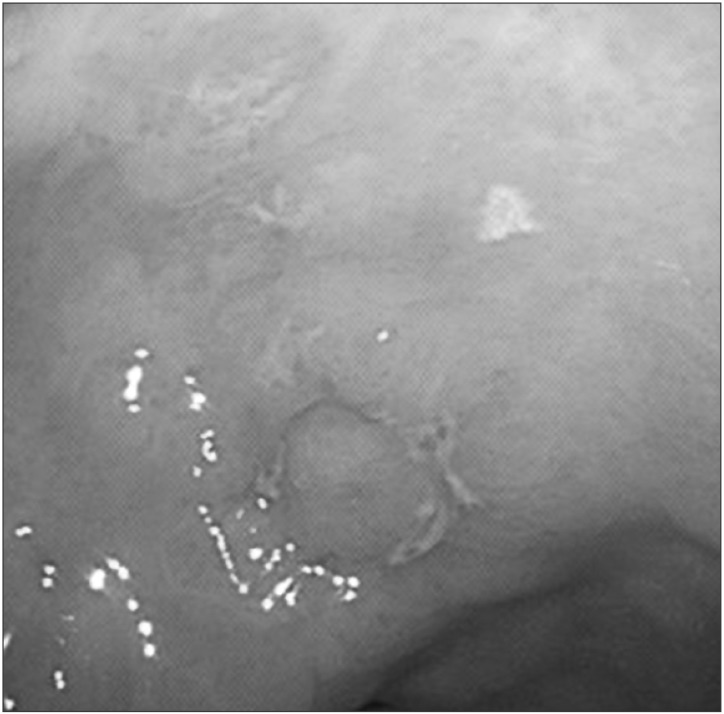 Esophagogastroduodenoscopy showing a small superficial elevated lesion in the antrum of the stomach.
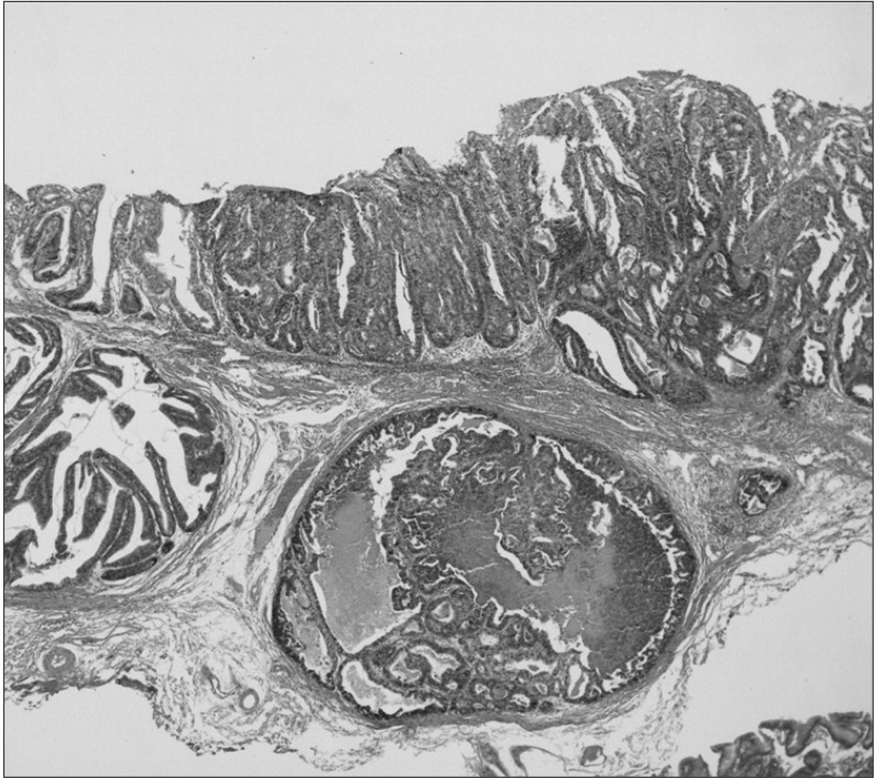 Histopathological examination of the specimens resected by endoscopic submucosal dissection revealing tubular adenocarcinoma with submucosal invasion (H&E, × 40).
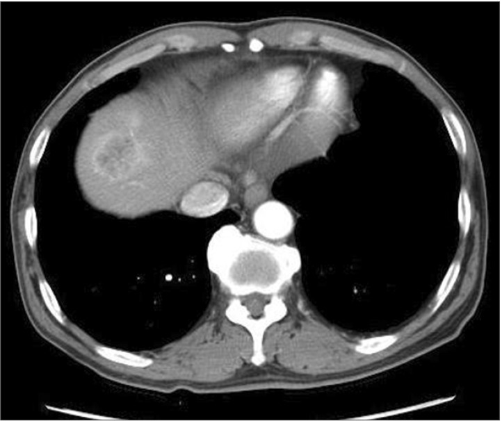 An abdominopelvic computed tomography scan showing a 3.5-cm-mass in the liver that demonstrated strong enhancement following the intravenous administration of contrast material.
본문에는 "1.7-cm superficially elevated (IIa)-type of EGC"로 씌여 있지만 EGC IIa+IIc라고 보는 것이 좋겠습니다. IIa는 깊은 경우가 드물지만 IIa+IIc는 깊은 경우가 많습니다. 전혀 다른 종류라고 보는 것이 옳습니다.
내시경 조직검사는 moderately differentiated adenocarcinoma였는데 ESD 병리는 moderately to poorly differentiated adenocarcinoma로 되어 있습니다. 아마도 mixed type이었던 것 아닌가 추정됩니다. Mixed type은 pure type 보다 좋지 못합니다.
이런 증례 보고는 흑백 사진이 아니라 천연색 사진으로 하였으면 더 좋았겠다고 생각됩니다. 종이 출력은 흑백으로 하더라도 인터넷에 올리는 자료는 천연색으로 하면 될 일입니다. 돈이 더 들어가는 것도 아니니까...
© 일원내시경교실 바른내시경연구소 이준행. EndoTODAY Endoscopy Learning Center. Lee Jun Haeng.