 EndoTODAY 내시경 교실
Beginner | ESA | Schedule | OPD
Seminars | Atlas | Recent | Links
![]() [위암 290 - 위암 대장 전이]
[위암 290 - 위암 대장 전이]
001 | 101 | 201 | 301 | 401 | 501 | 601 | 701 | 801 | 901 | 1000
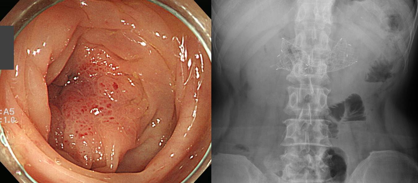
60대 여성입니다.

Advanced gastric carcinoma
1. Location : [1] lower third, [2] duodenum, Center at antrum and lesser curvature
2. Gross type : Borrmann type 3
3. Histologic type : tubular adenocarcinoma, poorly differentiated
4. Histologic type by Lauren : diffuse
5. Size : 9x8 cm
6. Depth of invasion : invades serosa (pT4a)
7. Resection margin: free from carcinoma
8. Lymph node metastasis : metastasis to 16 out of 28 regional lymph nodes (pN3b)
9. Lymphatic invasion : present
10. Venous invasion : not identified
11. Perineural invasion : present
12. Peritoneal cytology : negative
13. AJCC stage by 7th edition: T4a N3b
이 환자에서 사진이 흐린 것은 내시경 시스템의 contrast (= enhancement) 셋팅 문제였습니다. Contrast 버튼이 잘 못 눌려서 정상보다 낮게 설정되면 이렇게 뿌연 사진이 나옵니다. 내시경 제작회사에서 시스템 설계를 잘못한 탓이 큽니다만, 사용자도 주의할 필요가 있습니다. 우리나라에도 내시경 회사가 있었으면 좋겠습니다.
안타깝게도 몇 년 후 대장 폐쇄로 검사를 하였고 위암의 대장 전이로 진단하였으며 stent가 시술되었습니다.
* 참고: EndoTODAY 대장 전이
* 참고: EndoTODAY 위 전이
© 일원내시경교실 바른내시경연구소 이준행. EndoTODAY Endoscopy Learning Center. Lee Jun Haeng. (2015-7-28)