 EndoTODAY 내시경 교실
Beginner | ESA | Schedule | OPD
Seminars | Atlas | Recent | Links
 [위암 324]
001 | 101 | 201 | 301 | 401 | 501 | 601 | 701 | 801 | 901 | 1000
70대 중반 건진 내시경에서 경계가 불분명한 위암으로 의뢰되었습니다. ESD를 하였는데 piecemeal resection 되었고 lateral margin을 평가할 수 없는 상황이었습니다.
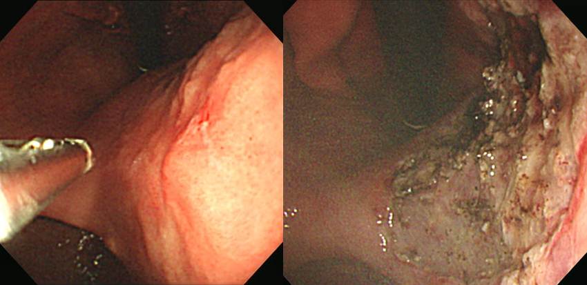
Endoscopic resection of early gastric carcinoma
1. Location : body, posterior wall
2. Gross type : EGC type IIa
3. Histologic type : tubular adenocarcinoma, moderately differentiated >>> tubular adenocarcinoma, poorly differentiated (3 %)
4. Histologic type by Lauren : intestinal
5. Size of carcinoma : (1) longest diameter, 24 mm (2) vertical diameter, 22 mm
6. Depth of invasion : invades mucosa (lamina propria) (pT1a)
7. Resection margin : (1) involved anterior and posterior resection margins, (2) proximal and distal resection margins cannot be determined due to piecemeal resection
8. Lymphatic invasion : not identified(N)
9. Venous invasion : not identified(N)
10. Perineural invasion : not identified(N)
11. Microscopic ulcer : absent
12. Histologic heterogeneity: present
연세, 24 mm lamina propria 암이었다는 점, 육안적으로는 residual tumor가 없었다는 점 등을 고려하여 수술과 경과관찰의 장단점을 잘 설명한 후 통상 재발률보다 높을 수 있지만 일단 주의깊게 경과관찰을 하기로 하였습니다. 그러나 아쉽게도 두번째 추적내시경 조직검사에서 암으로 확인하여 subtotal gastrectomy를 할 수 밖에 없었습니다.
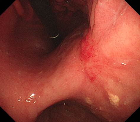 두번째 추적내시경. 암으로 확인됨.
Stomach, subtotal gastrectomy:
Early gastric carcinoma
1. Location : middle third, Center at angle and posterior wall
2. Gross type : EGC type IIc
3. Histologic type : tubular adenocarcinoma, moderately differentiated > tubular adenocarcinoma, poorly differentiated (30 %)
4. Histologic type by Lauren : intestinal
5. Size : 3x1.7 cm
6. Depth of invasion : invades mucosa (muscularis mucosae) (pT1a)
7. Resection margin: free from carcinoma
8. Lymph node metastasis : no metastasis in 34 regional lymph nodes (pN0)
9. Lymphatic invasion : not identified
10. Venous invasion : not identified
11. Perineural invasion : not identified
12. Peritoneal cytology : negative
13. AJCC stage by 7th edition: T1a, N0
Laterl margin involvement의 중요 원인 중 하나가 histological heterogeneity입니다. 순수한 것보다 섞인 것이 나쁩니다. 적어도 ESD 영역에서는...
© 2015-12-21. 일원내시경교실 바른내시경연구소 이준행