 EndoTODAY 내시경 교실
Beginner | ESA | Schedule | OPD
Seminars | Atlas | Recent | Links
 [위암 385]
001 | 101 | 201 | 301 | 401 | 501 | 601 | 701 | 801 | 901 | 1000
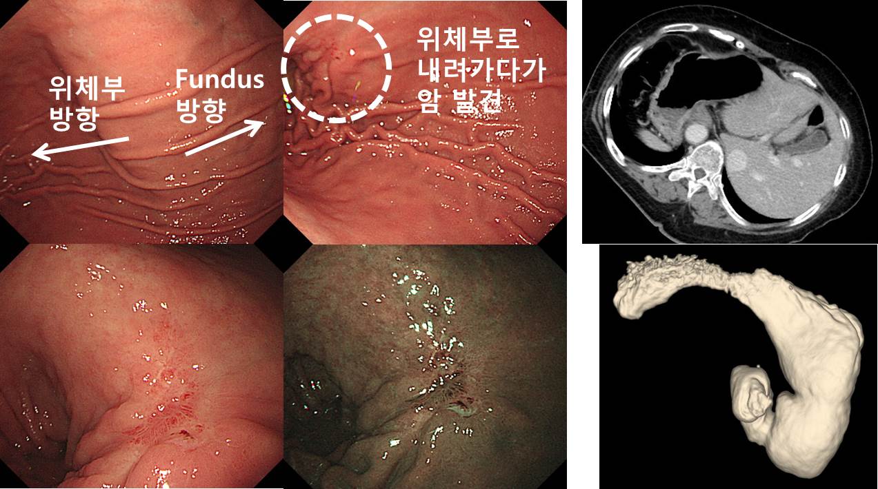
왜 이렇게 이상한 사진일까요? 방향이 영 이상합니다.
아래로
아래로
아래로
아래로
아래로
 Dextrocardia 환자에서 발견된 위암입니다. 식도에서 위로 들어간 다음 평소와 같은 우측 방향이 아니라 화면의 좌측방향으로 antrum으로 가는 길이 있었습니다. 위각부 직상부에서 암이 발견되었습니다.
Stomach, subtotal gastrectomy:
Early gastric carcinoma
1. Location : middle third, Center at low body and posterior wall
2. Gross type : EGC type IIb+IIc
3. Histologic type : tubular adenocarcinoma, poorly differentiated
4. Histologic type by Lauren : diffuse
5. Size : 3.3x3 cm
6. Depth of invasion : invades mucosa (muscularis mucosae) (pT1a)
7. Resection margin: free from carcinoma
8. Lymph node metastasis : no metastasis in 38 regional lymph nodes (pN0)
9. Lymphatic invasion : not identified
10. Venous invasion : not identified
11. Perineural invasion : not identified
12. Peritoneal cytology : negative
13. AJCC stage by 7th edition: pT1a N0
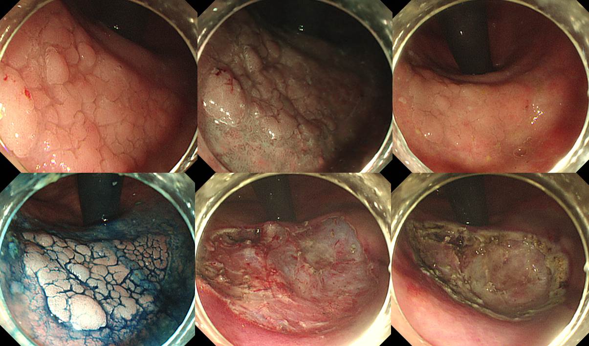
그런데 이 환자의 remnant stomach에서 adenoma가 발견되었습니다. (1) dextrocardia, (2) remnant stomach, (3) cardia 직하부, (4) 비교적 넓은 병소인지가 상당히 어려웠지만 ESD는 가능했습니다. 엄청 힘들었습니다. Resection margin이 부족할까봐 싶어서 360도 circumferential APC ablation을 추가했습니다.
* 참고: EndoTODAY situs inversus
© 2016-4-27. 일원내시경교실 바른내시경연구소 이준행