EndoTODAY 내시경 교실
Beginner | ESA | Schedule | OPD
Seminars | Atlas | Recent | Links
![]() [Gastric cancer 407]
[Gastric cancer 407]
001 | 101 | 201 | 301 | 401 | 501 | 601 | 701 | 801 | 901 | 1000
내시경 조직검사 결과 해석은 복잡할 수 있습니다. Tricky할 수 있습니다. 오래 된 증례 하나를 소개합니다. 할 때마다 결과가 달랐던 경우입니다.
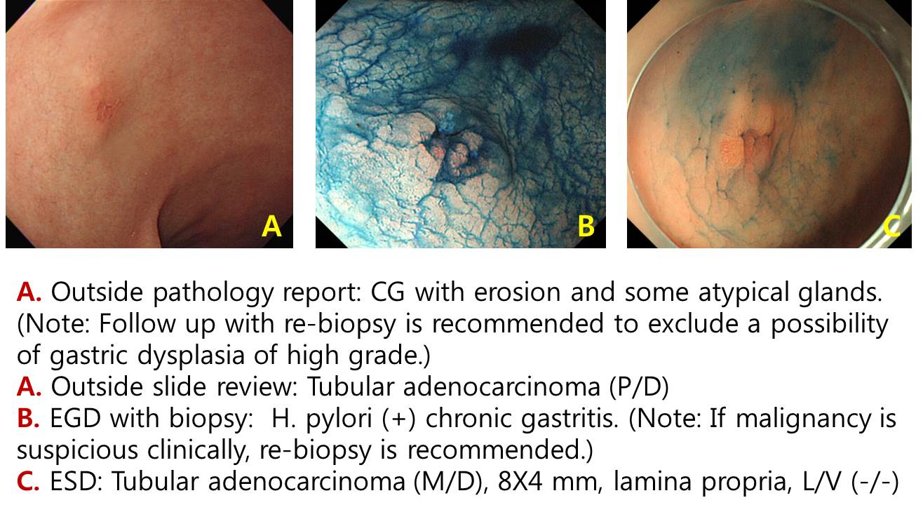
70대 남성입니다. "Chronic gastritis with erosion and some atypical glands. Note: Follow up with re-biopsy is recommended to exclude a possibility of gastric dysplasia of high grade."라는 병리결과를 가지고 방문하셨습니다. 내시경 사진은 아래와 같았습니다.
첫 외부 내시경
일단 ESD를 고려한 상태에서 외부 슬라이드 재판독을 하였는데, 의외로 "Tubular adenocarcinoma (P/D)"라고 보고되었습니다. 저는 poorly differentiated type 위암은 대부분 수술을 의뢰하고 있기 때문에, 바로 ESD를 하지 않고 내시경 재검을 하였습니다.
의뢰 후 내시경 재검
그런데 조직검사 결과가 "H. pylori (+) chronic gastritis. Note: If malignancy is suspicious clinically, re-biopsy is recommended."로 보고되었습니다. 여러분은 어떻게 하시겠습니까?
아래와 같이 설명하고 ESD를 권하였습니다. 돈 문제도 약간 설명하였습니다. (감독기관의 규정이 비현실적이고 비논리적이기 때문에 나름대로 약간의 방어성 설명도 하고 있습니다. 대한민국에서 의사를 하려면 필요한 일입니다. 치료는 잘 해드리고 돈 문제로 싸우는 경우가 많기 때문입니다. 왜 자꾸 어처구니 없는 삭감을 하는 것인지... 한심한 일입니다. )
외래 설명 (시술 전)
ESD는 평이하게 진행되었습니다.
ESD
최종 ESD 병리 결과는 아래와 같았습니다.
Stomach, endoscopic submucosal dissection:
Early gastric carcinoma
1. Location : antrum, anterior wall
2. Gross type : EGC type IIc
3. Histologic type : tubular adenocarcinoma, moderately differentiated
4. Histologic type by Lauren : intestinal
5. Size of carcinoma : (1) longest diameter, 8 mm (2) vertical diameter, 4 mm
6. Depth of invasion : invades mucosa (lamina propria) (pT1a)
7. Resection margin : free from carcinoma(N),safety margin : distal 10 mm, proximal 8 mm, anterior 12 mm, posterior 12 mm
8. Lymphatic invasion : not identified(N)
9. Venous invasion : not identified(N)
10. Perineural invasion : not identified(N)
11. Microscopic ulcer : absent
12. Histologic heterogeneity: absent
아래와 같이 설명하였습니다. 조기위암 내시경 치료 후 병리학적 완전절제가 되었을 때 설명하는 표준 형식입니다.
외래 설명 (시술 후)
다소 복잡하지만 요약하면 아래와 같습니다.
병리 결과 해석은 이렇게 tricky 할 수 있습니다. 많은 임상 경험이 중요합니다. 알파고가 할 수 있는 일이 아닙니다.
© EndoTODAY Endoscopy Learning Center. Jun Haeng Lee. 일원내시경교실 바른내시경연구소 이준행