 EndoTODAY 내시경 교실
Beginner | ESA | Schedule | OPD
Seminars | Atlas | Recent | Links
 [Gastric cancer 411]
001 | 101 | 201 | 301 | 401 | 501 | 601 | 701 | 801 | 901 | 1000
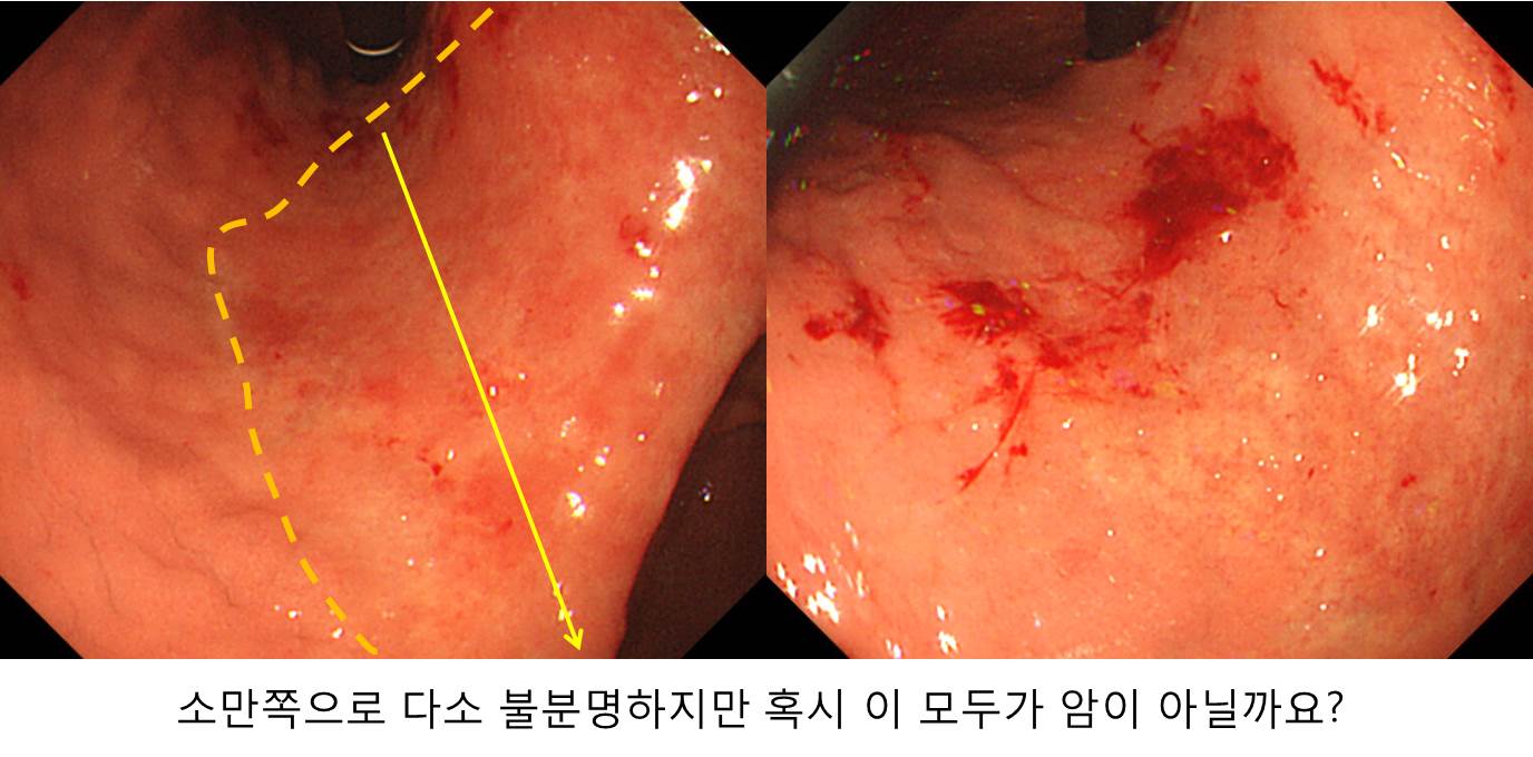
위각부 암으로 의뢰되었습니다. 수술전 내시경 재검을 하였는데 들문 직하부까지 위체부 소만을 따라서 위점막이 불규칙하였습니다. 위체상부 소만에서도 조직검사를 하였는데 그곳에서도 위암이 나왔습니다. Total gastrectomy를 하였고 아래와 같은 결과였습니다.
내시경 검사를 잘 해주신 선생님께 감사드립니다. 복잡한 일이 벌어질 뻔 하였습니다. 수술장에서 frozen 양성으로 절제범위를 조금씩 넓혔거나, subtotal gastrectomy 후 resection margin positive로 다시 한번 수술이 필요할 수 있었습니다. 질 좋은 내시경 검사가 환자에게 드리는 혜택은 대단합니다.
Early gastric carcinoma
1. Location : upper third and middle third, center at cardia, body and lesser curvature
2. Gross type : EGC type IIb+IIc
3. Histologic type : tubular adenocarcinoma, moderately(30%)+poorly (solid(50%) and poorly cohesive(20%)) differentiated
4. Histologic type by Lauren : mixed
5. Size : 10.5x6.3 cm
6. Depth of invasion : invades submucosa (sm2) (pT1b)
7. Resection margin: free from carcinoma, safety margin: proximal, 1.1 cm; distal, 4.3 cm
8. Lymph node metastasis : no metastasis in 67 regional lymph nodes (pN0)
9. Lymphatic invasion : present
10. Venous invasion : not identified
11. Perineural invasion : not identified
12. AJCC stage by 7th edition: T1b N0
© 일원내시경교실 바른내시경연구소 이준행. EndoTODAY Endoscopy Learning Center. Lee Jun Haeng.