EndoTODAY 내시경 교실
Beginner | ESA | Schedule | OPD
Seminars | Atlas | Recent | Links
![]() [Gastric cancer 439 - tumor island]
[Gastric cancer 439 - tumor island]
001 | 101 | 201 | 301 | 401 | 501 | 601 | 701 | 801 | 901 | 1000
내시경 초심자로부터 이런 질문을 받았습니다. "아래 그림을 자주 보게 되는데요... Fold, edge, margin, base는 대강 알겠는데 tumor island는 무엇인지 궁금합니다. Atlas에도 잘 나와 있지 않았습니다."
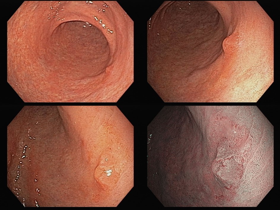
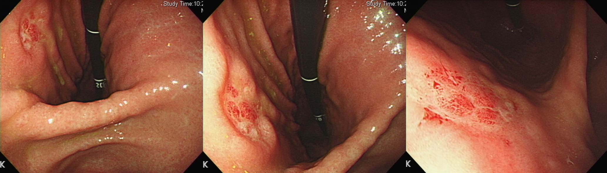
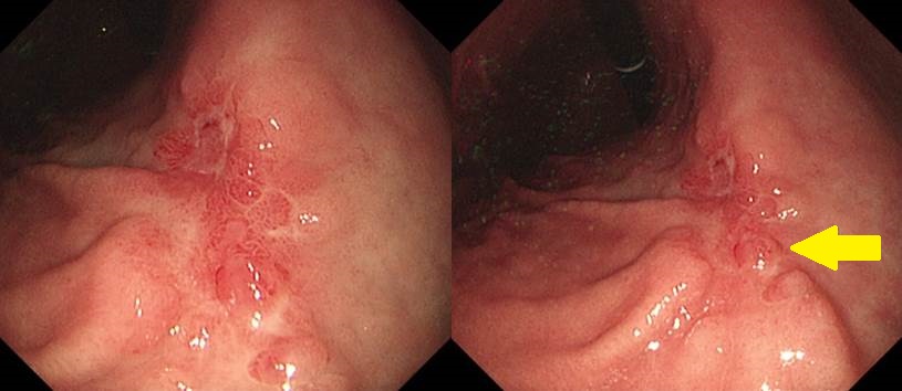
함몰형 위암의 전형적 abnormal folds. Poorly differentiated tubular adenocarcinoma. 처음부터 복수와 bilateral ovarian metastasis가 있었음.
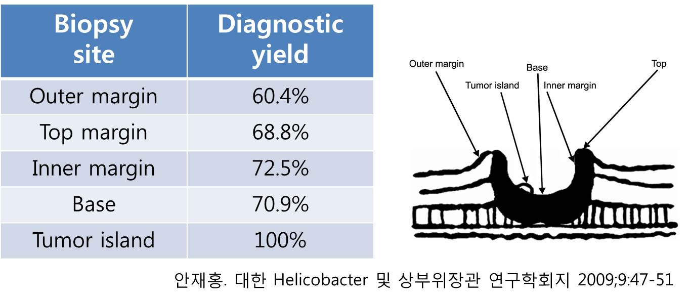
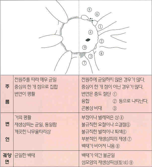
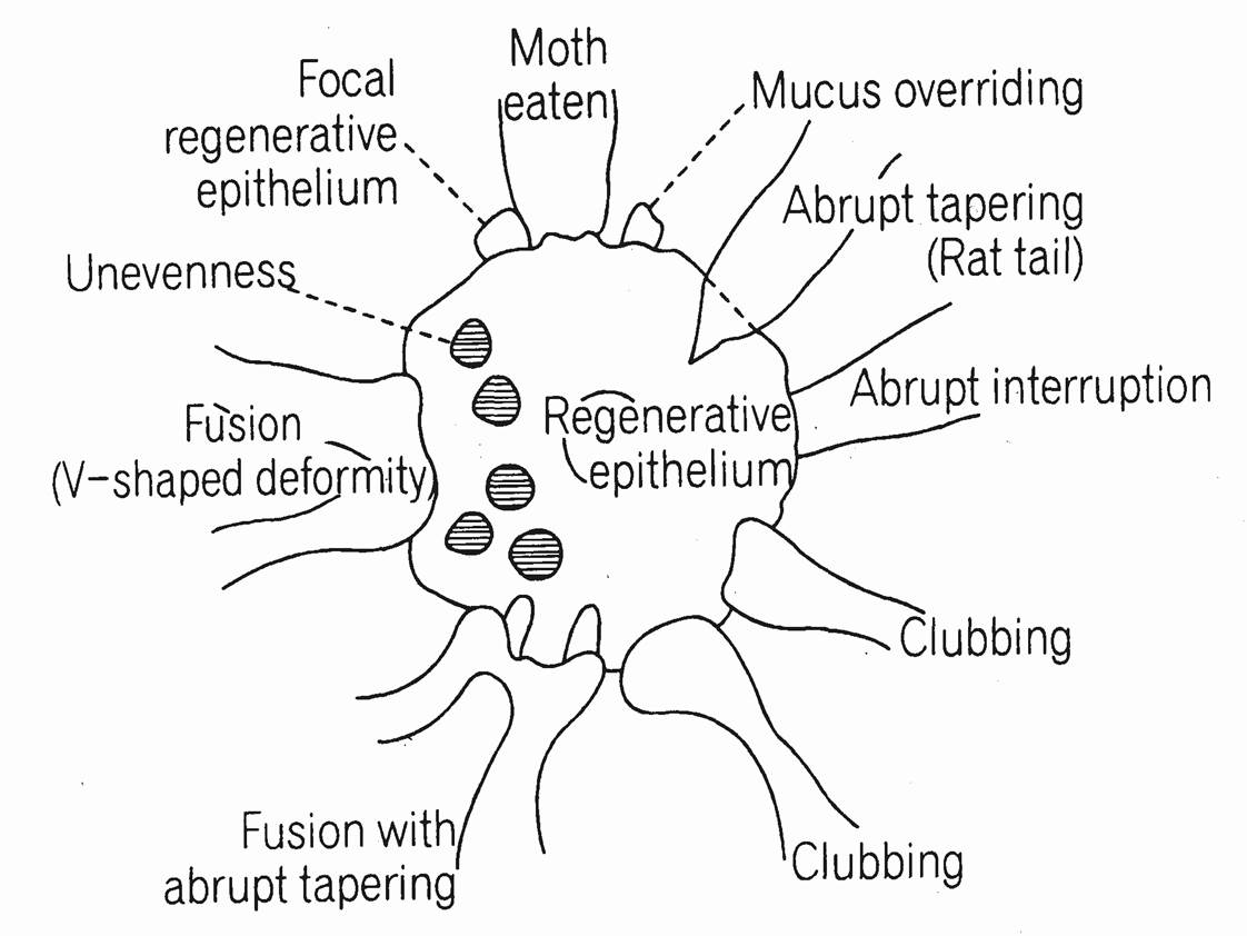
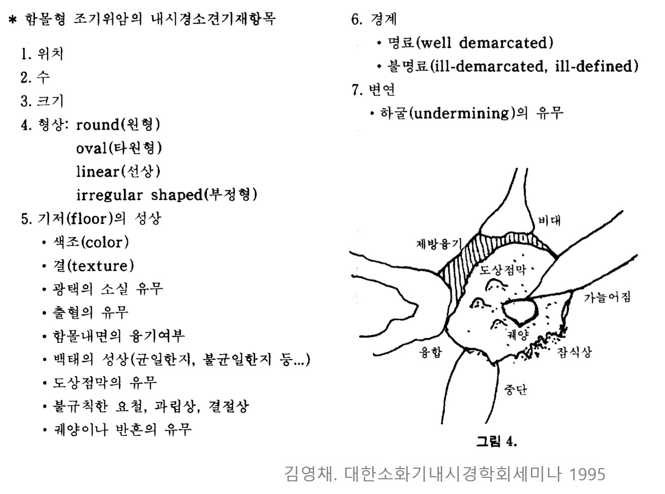
Tumor island는 궤양 바닥에서 돌출된 부위입니다. 이름 그대로 섬(island)입니다. Tumor island는 양성 위궤양과 함몰형 위암의 감별진단에도 요긴한 개념이고, 조직검사 target으로도 유용합니다. 아래는 forcep biopsy 위치에 따른 암 양성률을 조사한 서울아산병원의 연구입니다. 함몰형 병소의 부위별 암 진단율을 분석한 것인데, tumor island에서는 암 양성률이 100%였습니다. 그러나 outer margin, top margin, inner margin, base의 암 진단율은 생각만큼 큰 차이가 없었습니다.
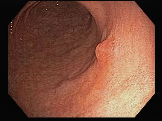
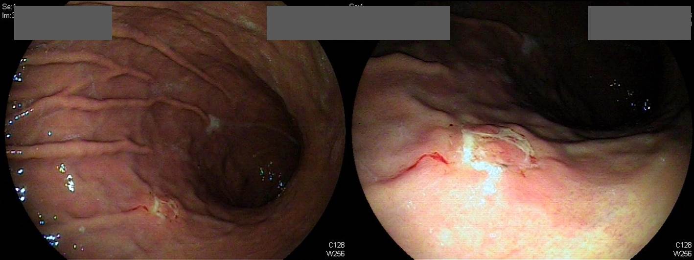
우선 가장 전형적인 tumor island를 보인 위암 증례 세 개를 소개합니다.
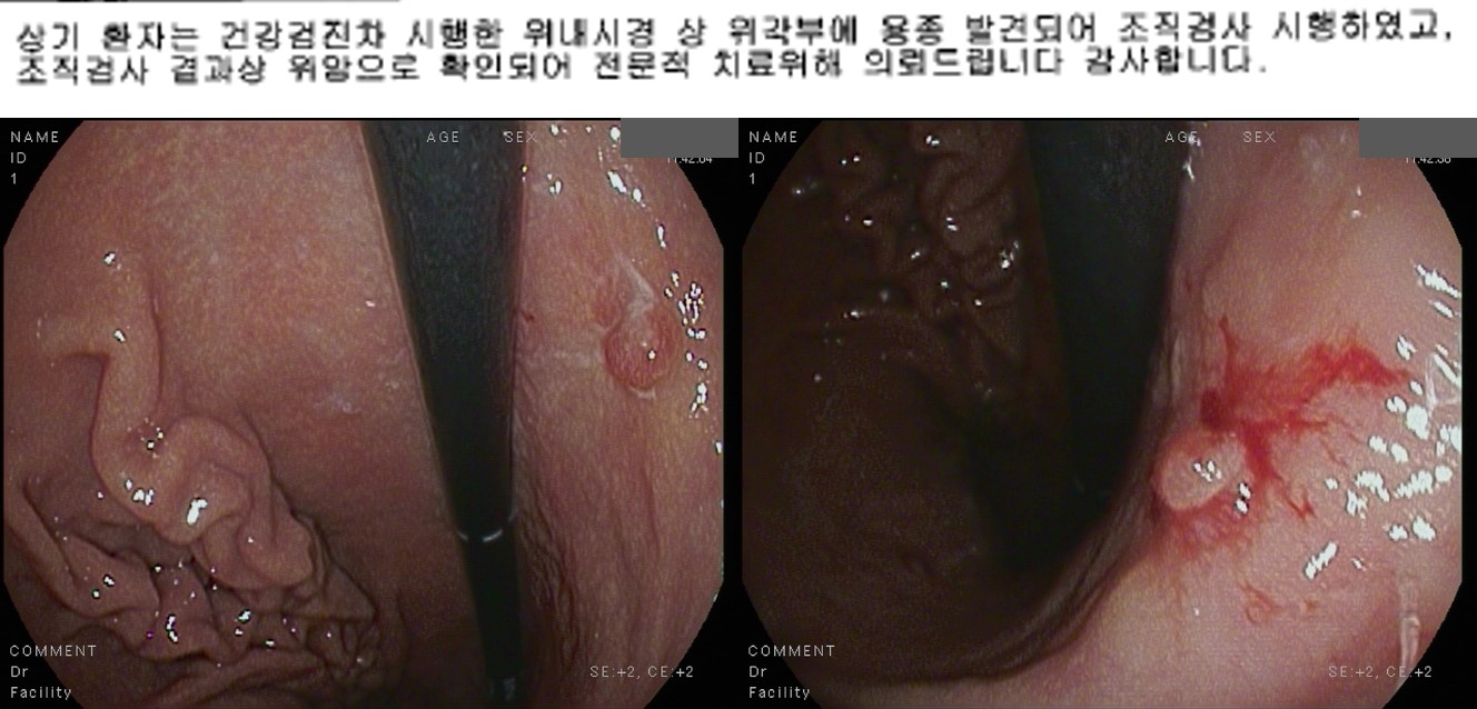
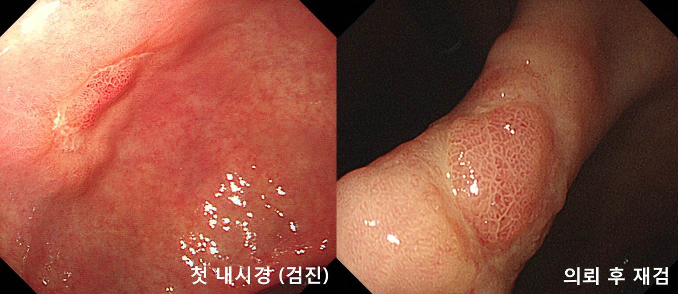
멋진 사진입니다. 처음 발견하신 분께서 전형적인 tumor island를 보고 조직검사 전 chromoendoscopy 사진을 남기셨습니다. 감사합니다.
Early gastric carcinoma
1. Location : lower third, Center at antrum and greater curvature
2. Gross type : EGC type IIb
3. Histologic type : signet-ring cell carcinoma
4. Histologic type by Lauren : diffuse
5. Size : 1.5x0.6 cm
6. Depth of invasion : invades mucosa (lamina propria) (pT1a)
7. Resection margin: free from carcinoma, safety margin: proximal 9.5 cm, distal 5.4 cm
8. Lymph node metastasis : no metastasis in 42 regional lymph nodes (pN0)
9. Lymphatic invasion : not identified
10. Venous invasion : not identified
11. Perineural invasion : not identified
12. Peritoneal cytology : negative
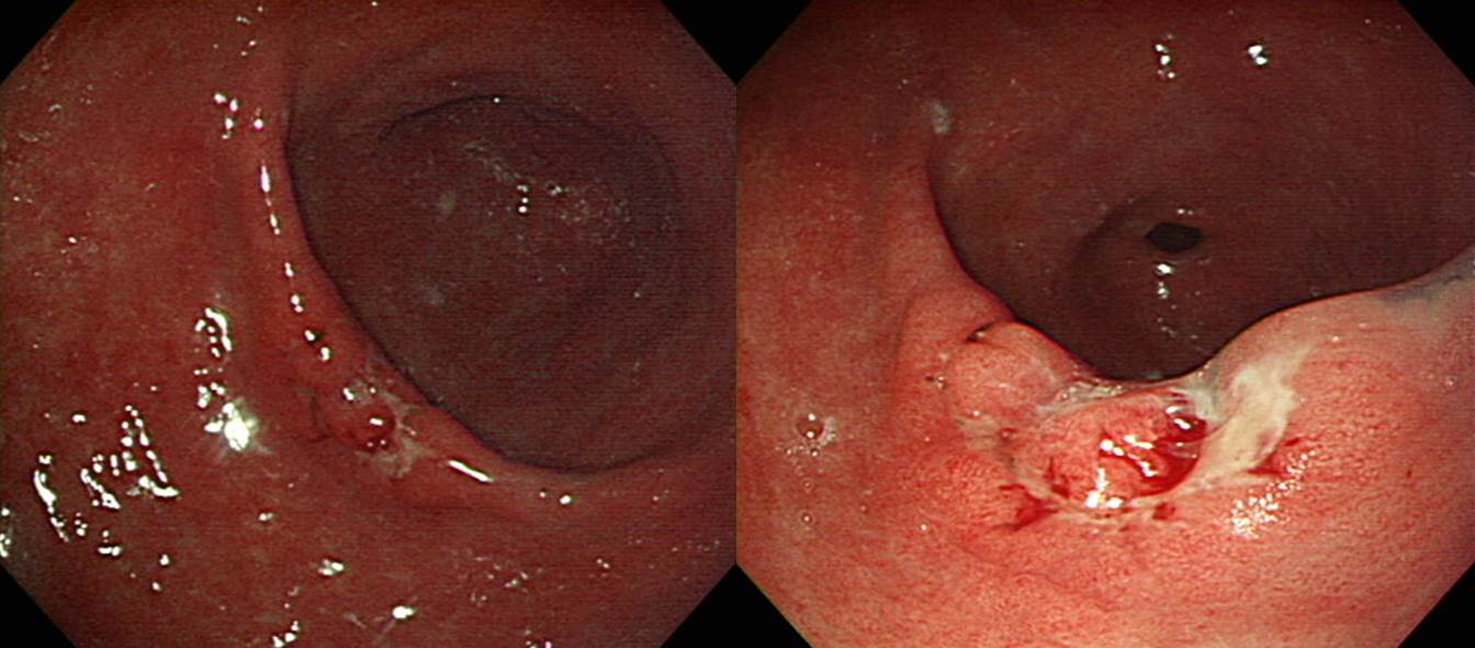
전형적인 tumor island
환자가 수술을 거부하여 수술하지 못한 경우입니다.
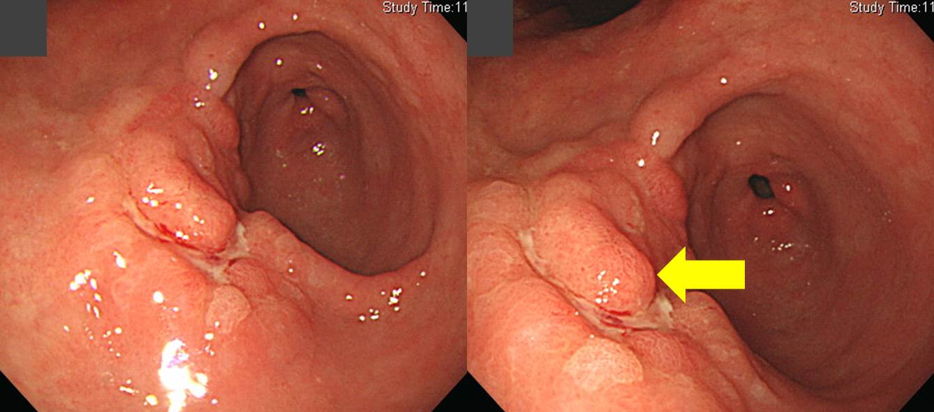
일부 증례는 tumor island 같기는 했지만 그 높이가 너무 낮아서 '함몰부 바닥의 발적'으로 표현해도 좋을 것 같은 경우도 있었습니다.
Early gastric carcinoma
1. Location : middle third, Center at low body and greater curvature
2. Gross type : EGC type IIb+IIc
3. Histologic type : signet-ring cell carcinoma
4. Histologic type by Lauren : diffuse
5. Size : 1.7x1.4 cm
6. Depth of invasion : invades mucosa (lamina propria) (pT1a)
7. Resection margin: free from carcinoma, safety margin: proximal 3 cm, distal 6.8 cm
8. Lymph node metastasis : no metastasis in 26 regional lymph nodes (pN0)
9. Lymphatic invasion : not identified
10. Venous invasion : not identified
11. Perineural invasion : not identified
외부 조직검사에서 moderate dysplasia로 오셨으나 최종 결론은 SM cancer였습니다.
Early gastric carcinoma
1. Location : lower third, center at antrum and posterior wall
2. Gross type : EGC type IIa+IIc
3. Histologic type : tubular adenocarcinoma, moderately differentiated
4. Histologic type by Lauren : intestinal
5. Size : 2.5x2cm
6. Depth of invasion : extension to submucosa (sm2) (pT1b)
7. Resection margin: free from carcinoma, safety margin: proximal, 8.2 cm; distal, 3.5 cm
8. Lymph node metastasis : no metastasis in 38 regional lymph nodes (pN0)
9. Lymphatic invasion : not identified
10.Venous invasion : not identified
11.Perineural invasion : not identified
[More cases with tumor island]
Early gastric carcinoma
1. Location : middle third, Center at body and lesser curvature
2. Gross type : EGC type IIc
3. Histologic type : tubular adenocarcinoma, poorly solid(50%) and poorly cohesive(50%)
4. Histologic type by Lauren : mixed
5. Size : 2.3x1.2 cm
6. Depth of invasion : invades mucosa (muscularis mucosae) (pT1a)
7. Resection margin: free from carcinoma, safety margin: proximal 5.4 cm, distal 3.6 cm
8. Lymph node metastasis : no metastasis in 27 regional lymph nodes (pN0)
9. Lymphatic invasion : not identified
10. Venous invasion : not identified
11. Perineural invasion : not identified
12. AJCC stage by 7th edition: pT1a N0
Advanced gastric carcinoma
1. Location : lower third, Center at antrum and lesser curvature
2. Gross type : Borrmann type 2
3. Histologic type : tubular adenocarcinoma, moderately differentiated
4. Histologic type by Lauren : intestinal
5. Size : 5x2.7 cm
6. Depth of invasion : invades muscularis propria (pT2)
7. Resection margin: free from carcinoma
8. Lymph node metastasis : metastasis to 1 out of 22 regional lymph nodes (pN1) (1/22: "1", 0/2; "3", 0/5; "4", 1/6; "4sb", 0/0; "5", 0/0; "6", 0/2; "8a", 0/2; "7", 0/2; "9", 0/1; "11p", 0/0; "12a", 0/2)
9. Lymphatic invasion : not identified
10. Venous invasion : not identified
11. Perineural invasion : not identified
12. Peritoneal cytology : negative

Early gastric carcinoma
1. Location : lower third, center at antrum and lesser curvature
2. Gross type : EGC type IIc
3. Histologic type : tubular adenocarcinoma, poorly differentiated
4. Histologic type by Lauren : diffuse
5. Size : 5.4x2.6 cm
6. Depth of invasion : extension to submucosa (sm3) (pT1b)
7. Resection margin: free from carcinoma, safety margin: proximal, 5.5 cm; distal, 3.5 cm
8. Lymph node metastasis : no metastasis in 48 regional lymph nodes (pN0)
9. Lymphatic invasion : not identified
10.Venous invasion : not identified
11.Perineural invasion : not identified
Advanced gastric cancer
1. Location : middle third, Center at low body and anterior wall
2. Gross type : Borrmann type 2
3. Histologic type : signet-ring cell carcinoma
4. Histologic type by Lauren : mixed
5. Size : 2.0x1.7 cm
6. Depth of invasion : invades muscularis propria (pT2)
7. Resection margin: free from carcinoma, safety margin: proximal 3 cm, distal 13.5 cm
8. Lymph node metastasis : no metastasis in 19 regional lymph nodes (pN0)
9. Lymphatic invasion : present
10. Venous invasion : present(intramural)
11. Perineural invasion : present
12. Peritoneal cytology : atypical cells
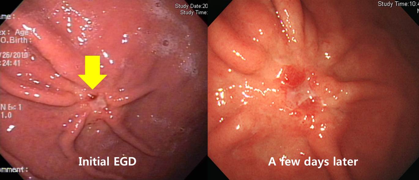
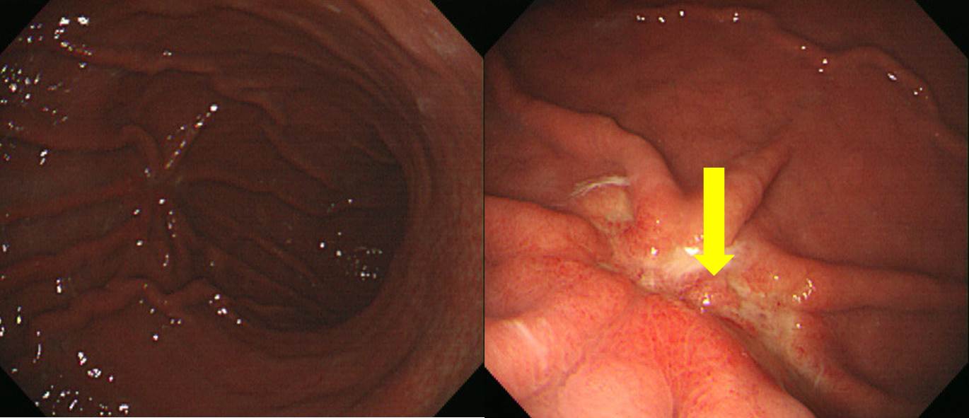
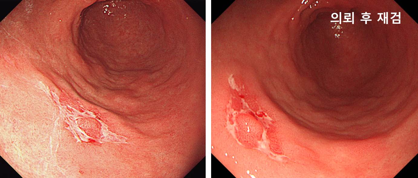
틀림없이 조기위암입니다. 함몰형인데 중앙에 융기부(tumor island)가 있는 상태로 발견되었습니다. 조직검사로 위암이 확인되어 의뢰되었고 재검을 하였는데, 첫 검사에서 보였던 tumor island가 없어진 모양이었습니다. 조직검사로 tumor island가 사라진 것 같습니다.
Stomach, radical subtotal gastrectomy:
Early gastric carcinoma
1. Location : lower third, Center at antrum and greater curvature
2. Gross type : EGC type IIc
3. Histologic type : tubular adenocarcinoma, moderately differentiated
4. Histologic type by Lauren : intestinal
5. Size : 2.3x0.8 cm
6. Depth of invasion : invades submucosa (sm1) (pT1b)
7. Resection margin: free from carcinoma, safety margin: proximal 11 cm, distal 2.5 cm
8. Lymph node metastasis : no metastasis in 51 regional lymph nodes (pN0)
9. Lymphatic invasion : not identified
10. Venous invasion : not identified
11. Perineural invasion : not identified

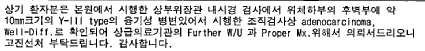
signet ring cell carcinoma, 1.3cm, SM2
Signet ring cell carcinoma. 한 병원에서 ESD 권유받고 second opinion 위하여 오신 분입니다. 당연히 수술을 권했습니다.

Stomach, LC of angle, ESD: Early gastric carcinoma
1. Location : angle, lesser curvature
2. Gross type : EGC type IIc
3. Histologic type : tubular adenocarcinoma, well differentiated
4. Histologic type by Lauren : intestinal
5. Size of carcinoma : (1) longest diameter, 36 mm (2) vertical diameter, 23 mm
6. Depth of invasion : invades mucosa (muscularis mucosa) (pT1a)
7. Resection margin : free from carcinoma(N), safety margin : distal 6 mm, proximal 3 mm, anterior 8 mm, posterior 8 mm, deep 500 ㎛
8. Lymphatic invasion : not identified(N)
9. Venous invasion : not identified(N)
10. Perineural invasion : not identified(N)
11. Microscopic ulcer : absent
12. Histologic heterogeneity: absent
[EndoTODAY gastric cancer 833]
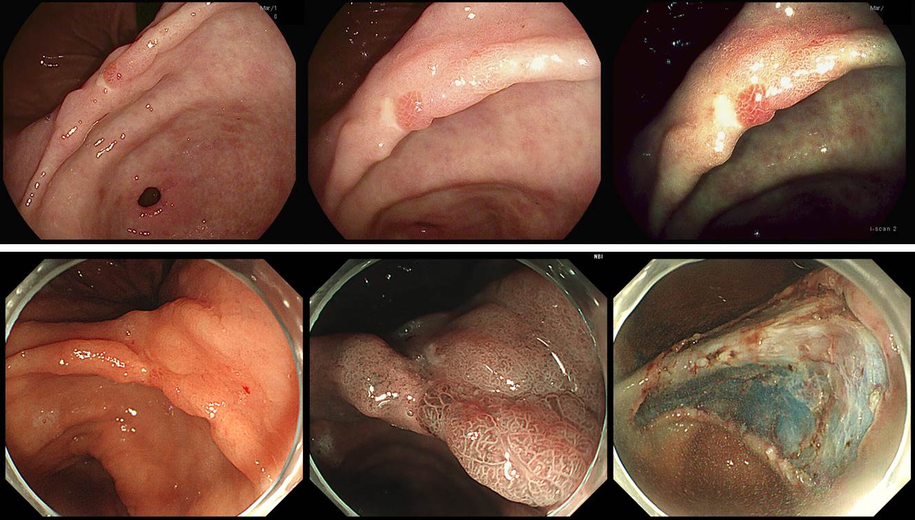
A patient with a gastric polyp, which was found in the screening endoscopy, was referred for the endoscopic treatment.
When I reviewed the outside endoscopic images, it was not a polypoid EGC, but a depressed type EGC with a small elevated lesion (tumor island). Endoscopic resection was done as usual.

ESD: Early gastric carcinoma
1. Location : angle, posterior wall
2. Gross type : EGC type IIc
3. Histologic type : tubular adenocarcinoma, moderately differentiated
4. Histologic type by Lauren : intestinal
5. Size of carcinoma : (1) longest diameter, 18 mm (2) vertical diameter, 14 mm
6. Depth of invasion : invades mucosa (lamina propria) (pT1a)
7. Resection margin : free from carcinoma(N), safety margin : distal 11 mm, proximal 4 mm, anterior 12 mm, posterior 6 mm, deep 300 ㎛
8. Lymphatic invasion : not identified(N)
9. Venous invasion : not identified(N)
10. Perineural invasion : not identified(N)
11. Pre-existing adenoma : none
12. Microscopic ulcer : absent
13. Histologic heterogeneity: absent
It is easier to find elevated lesions than depressed lesions. When a lesion was found, we need to see the surrounding mucosa carefully.
[EndoTODAY gastric cancer 853]
A patient with a small EGC was referred. It was described as a 10mm elevated lesion (biopsy: well differentiated adenocarcinoma).
When I reviewed more images, it was a depressed type EGC with a small elevated lesion (tumor island).
ESD was done.
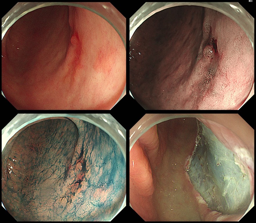
ESD: Early gastric carcinoma
1. Location : antrum, posterior wall
2. Gross type : EGC type IIc
3. Histologic type : tubular adenocarcinoma, moderately differentiated
4. Histologic type by Lauren : intestinal
5. Size of carcinoma : (1) longest diameter, 16 mm (2) vertical diameter, 14 mm
6. Depth of invasion : invades mucosa (muscularis mucosa) (pT1a)
7. Resection margin : free from carcinoma(N), safety margin : distal 10 mm, proximal 10 mm, anterior 10 mm, posterior 12 mm, deep 400 ㎛
8. Lymphatic invasion : not identified(N)
9. Venous invasion : not identified(N)
10. Perineural invasion : not identified(N)
11. Microscopic ulcer : absent
12. Histologic heterogeneity: absent
© 일원내시경교실 바른내시경연구소 이준행. EndoTODAY Endoscopy Learning Center. Lee Jun Haeng.