![]() [Description exercise 2 ЧиМГ] - №ћ
[Description exercise 2 ЧиМГ] - №ћ
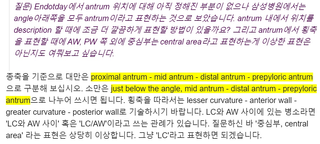
УтЧїМК РЇПАРЬЖѓДТ ИЛРК АЁБоРћ ОВСі ИПНУДй. (ПЯРќШї РЬСиЧр АГРЮ Л§АЂРдДЯДйИИ) RT-induced hemorrhagic gastritisГЊ immune checkpoint inhibitor-induced hemorrhagic gastritisПЭ ААРЬ ЦЏМіЧб АцПьИІ ЛЉАэ УтЧїМК РЇПАРЬЖѓДТ ИЛРЛ ОВСі ОЪЕЕЗЯ БЧЧеДЯДй.
РћР§Чб ГЛНУАц АЫЛч НУАЃРК Ию КаРЯБюПф? РњДТ РЇГЛНУАцРК РќУМ АЫЛчНУАЃ 5Ка, ДыРхГЛНУАцРК withdrawal time 6КаРЬЖѓАэ Л§АЂЧеДЯДй. ГЛНУАцРЛ УГРН НУРлЧЯДТ 1ГтТї fellowРЧ АцПь ПРРќ РЇГЛНУАц 12ИэРЬ РћДчЧеДЯДй. АЫЛч НУАЃРК 5КаРЬСіИИ sedation, ШЏРкЦФОЧ, УГЙц, АсАњРдЗТ ЕюРЧ НУАЃЕЕ ЧЪПфЧЯБт ЖЇЙЎРдДЯДй. 1ГтТї СпЙн ШЄРК 2ГтТї РЬЛѓРЬ ЕЧИщ СЛ Дѕ ИЙРК АЫЛчИІ Чв Мі РжНРДЯДй. АЫЛчИІ Рп Чи ЕхЗСОпАкДйДТ Л§АЂРИЗЮ Чб ИэРЧ ШЏРкПЁМ ГЪЙЋ Бф НУАЃРЛ ОВДТ АЭРК ШЏРкПЁАдЕЕ ЧиАЁ ЕЩ Мі РжНРДЯДй. ЦЏШї КёСјСЄ ГЛНУАцРЧ АцПьДТ ДѕПэ БзЗЏЧеДЯДй. ЙЙЕчСі РћР§Чб АЭРЬ ССНРДЯДй. ГЪЙЋ КќИЃСіЕЕ ОЪАэ ГЪЙЋ ДРИЎСіЕЕ ОЪАд. ёщщМ.
DEX 2ПЁ ДыЧб ПЉЗЏ СњЙЎРЬ РжОюМ ДфКЏ ЕППЕЛѓРЛ ИИЕщОю КИОвНРДЯДй.
2025-1-9.
![]() Case 8
Case 8
МвАп: Squamocolumnar junctionРЬ diaphragmatic orficeКИДй 2cm РЬЛѓ ПУЖѓПЭ РжРН. ЧЯКЮ НФЕЕПЁ 4АГ СЄЕЕРЧ linearЧЯАэ ОЦЗЁТЪРИЗЮ АЅМіЗЯ ГаОюСіДТ exudateЗЮ ЕЄШљ mucosal breakАЁ РжРИИч ИЧ ЧЯДмПЁМ circumferenceРЧ 75% РЬЛѓ fusionЕЧОю РжРН.
СјДм: (1) Sliding hiatal hernia, (2) Reflux esophagitis, LA-D
[РЬСиЧр comment]
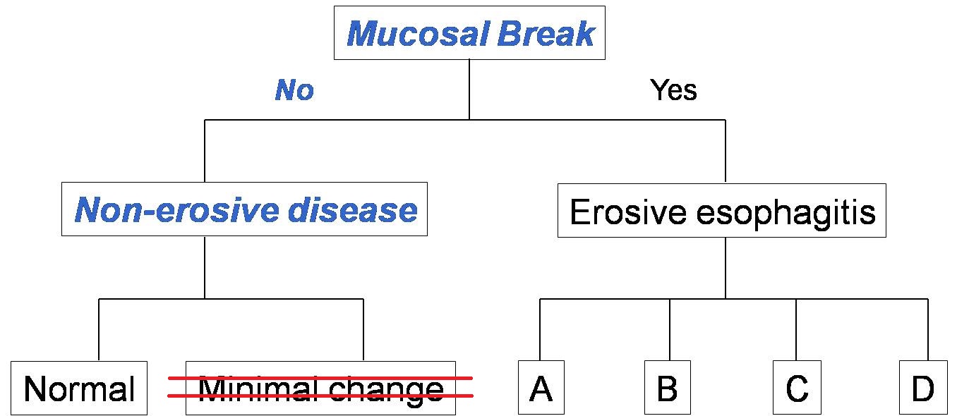
СјДмИэРЛ GERDЗЮ СжНХ КаРЬ ИЙОвНРДЯДй. GERDДТ РгЛѓ СјДмИэРдДЯДй. GERDДТ ГЛНУАцПЁМ mucosal break(ДыА erosionРЬГЊ ulcerИІ ИЛЧЯДТ АЭРг)АЁ ЖбЗЧЧб reflux esophagitis (АќЗЪРћРИЗЮ reflux esophagitis = erosive esophagitis)ПЭ mucosal breakАЁ ОјДТ non-erosive reflux disease (NERD)ИІ ЦїЧдЧЯДТ АГГфРдДЯДй (GERD = reflux esophagitis + NERD). ЕћЖѓМ РќЧќРћРЮ ЛъПЊЗљ СѕЛѓРЬ РжДТ ШЏРкПЁМ ГЛНУАц СјДмРК mucosal breakАЁ РжРИИщ reflux esophagitis LA-A/B/C/DЗЮ, mucosal breakАЁ ОјРИИщ СЄЛѓРИЗЮ ГЛИщ ЕЧАкНРДЯДй. Hiatal herniaАЁ РжРИИщ КДБтЧи СжНЪНУПР. ГЛНУАц СјДмИэРИЗЮ GERDИІ ОВСі ИЖНУБт ЙйЖјДЯДй.
СјДмИэРИЗЮ "LES incompetence"РЬЖѓ ОВНХ КаЕЕ АшМЬНРДЯДй. ГЛНУАц СјДмРК anatomical diagnosisРдДЯДй. ЙЋИЎЧЯПЉ functional diagnosisИІ КйРЬИщ РћСі ОЪРК ШЅМБРЬ ЙпЛ§ЧеДЯДй. LES incompetence ДыНХ hiatal herniaАЁ РћР§Чб СјДмИэРдДЯДй.
РЇНФЕЕПЊЗљСњШЏРЧ СјДм Йз ФЁЗсПЁ ДыЧб ОЦЗЁ ЕППЕЛѓРЛ ВР КИНУБт ЙйЖјДЯДй.

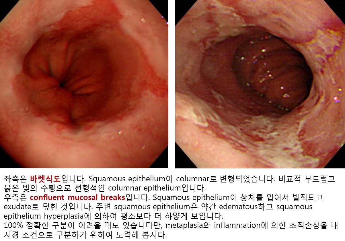
ПЊЗљМК НФЕЕПАПЁ РЧЧб mucosal breakАЁ КИХыРЧ АцПьУГЗГ АЁДУСі ОЪАэ ГаРК И№ОчРЬОњРИЙЧЗЮ АЃШЄ ЙйЗПНФЕЕЗЮ ДфЧб КаРЬ АшМЬНРДЯДй. БзЗЏГЊ ЙйЗПНФЕЕДТ ПАСѕ МвАпКИДйДТ metaplasiaРдДЯДй. БзГЩ ПЙЛл columnar colored mucosaЗЮ КИРЬЙЧЗЮ ПЕ ДйИЈДЯДй. ОЦЗЁИІ ТќАэЧЯММПф.
РЬ СЄЕЕ НЩЧб ПЊЗљМКНФЕЕПАПЁДТ ДУ hiatal herniaАЁ ЕПЙнЕЧБт ИЖЗУРдДЯДй. Hiatal herniaДТ ГЛНУАц УЪНЩРкЕщРЬ ПУЙйИЅ АГГфРЛ РтБтАЁ ОюЗСПю КЮКаРЬБтЕЕ ЧеДЯДй. ОЦЗЁ ЕППЕЛѓРЛ ВР КИНУБт ЙйЖјДЯДй.
Hiatal herniaРЧ КаЗљИІ mixed paraesophageal ЧќРИЗЮ ДфЧб КаРЬ АшМЬНРДЯДй. РЇПЁМ retroflexionЧб ЛчСјРЬ ОјОюМ СЄШЎШї ОЫ Мі ОјСіИИ РњДТ sliding herniaЗЮ КИОвДТЕЅПф... ЛчНЧ mixed paraesophageal herniaРЯ АЁДЩМКЕЕ ОјСі ОЪНРДЯДй.
СжКЏ СЁИЗРЛ О№БоЧЯНХ КаРЬ АшМХМ ДфКЏРЛ ЕхЗШНРДЯДй. ДйМв И№ШЃЧЯАд ДфЧпНРДЯДй.
2020Гт КЛАњ 4ЧаГт ЧаЛ§РЧ СњЙЎПЁ ДфЧпНРДЯДй.
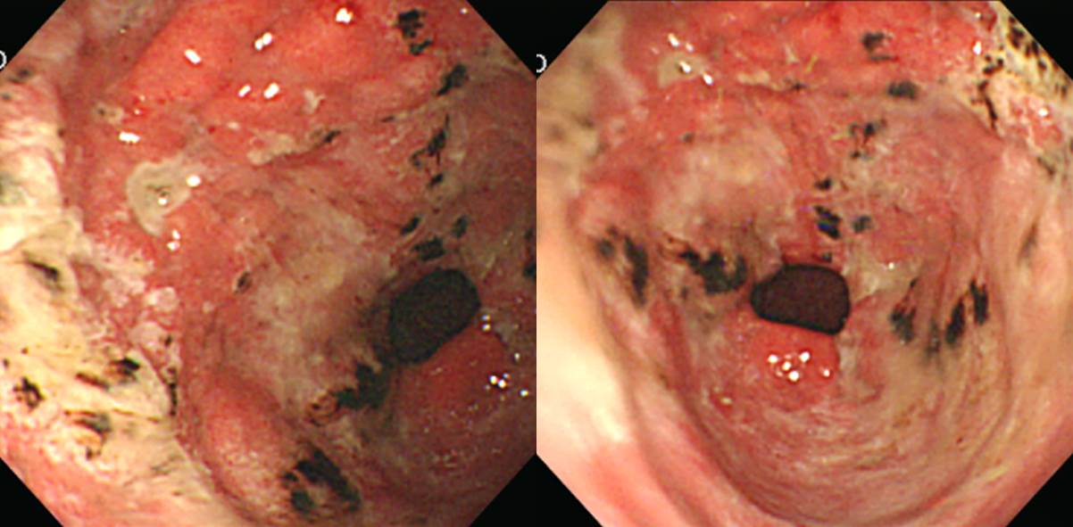
![]() Case 9. Midesophagus.
Case 9. Midesophagus.
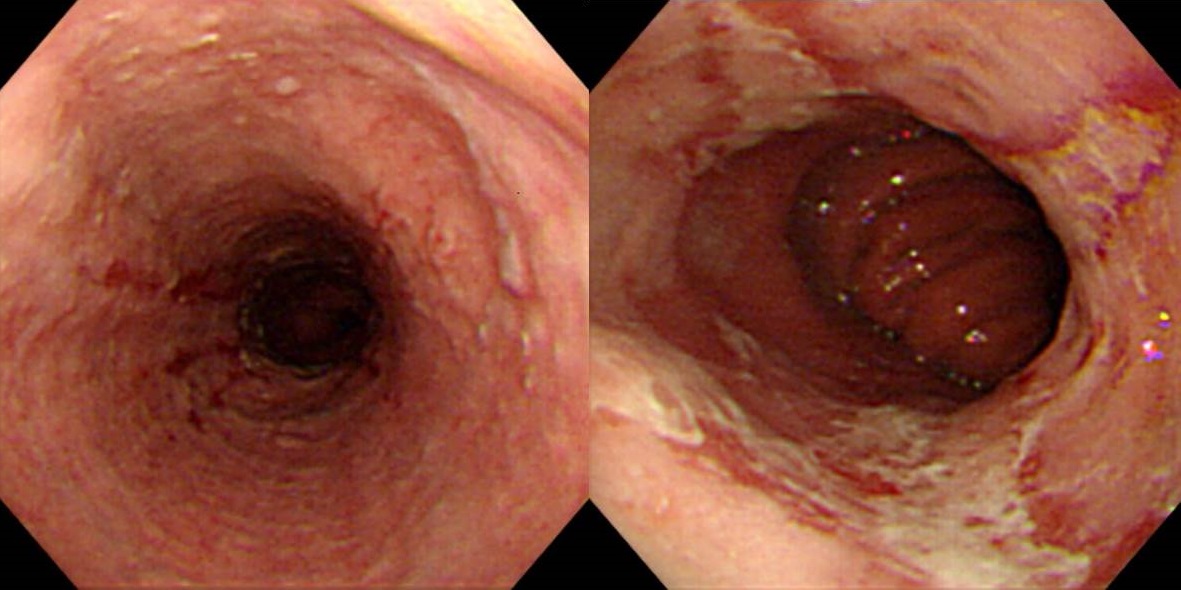
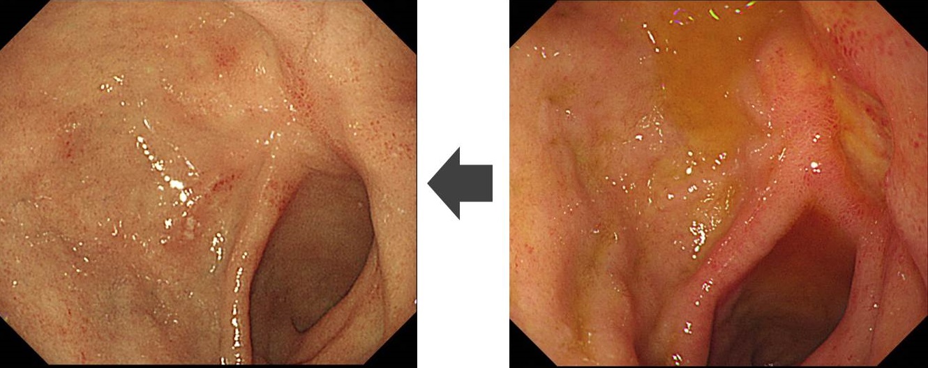
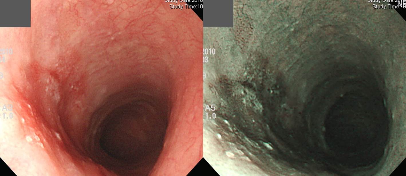 left: white light endoscopy picture, right: NBI (narrow band imaging) picture
МвАп: СпКЮНФЕЕ 6НУ~9НУ ЙцЧтПЁ 2 cm АЁЗЎРЧ slightly depressed hyperemic areaАЁ РжРН. 9НУ ЙцЧтРИЗЮ 0.5cm СЄЕЕРЧ АцЙЬЧб РЖБтКЮЕЕ КИРг. АцАшДТ ОрАЃ КвБдФЂЧЯСіИИ РќУМРћРИЗЮ ЕеБй КДМвРЬАэ, ЧЅИщРК РЯКЮ irregularЧб ОчЛѓРЬИч РЯКЮ white papule ОчЛѓРЧ exudateАЁ КйОю РжРН.
СјДм: Superficial esophageal cancer, IIb - ТќАэ: IIcЖѓАэ ЧиЕЕ ЙЋГЧв АЭ ААНРДЯДй.
[РЬСиЧр comment]
НФЕЕРЧ СіИЇРК ДыА 2.5 cm РдДЯДй. РЬИІ АэЗСЧЯПЉ ХЉБтИІ СќРлЧи СжНЪНУПР. РњДТ flat ШЄРК depressedЗЮ Л§АЂЧЯПДДТЕЅ РЧПмЗЮ ПЉЗЏКаРЬ slightly elevatedЗЮ КИМЬБКПф.^^ АќЗЪПЁ ЕћЖѓ НФЕЕПЁМДТ ГЛНУАц СјДмПЁ early esophageal cancerЖѓАэ ОВСі ИЛАэ superficial esophageal cancerЖѓАэ ОВНУБт ЙйЖјДЯДй. 'АцАшАЁ И№ШЃЧЯДй'Аэ ЧЅЧіЧв СЄЕЕЗЮ И№ШЃЧЯСі ОЪРКЕЅПф... РЬ СЄЕЕДТ КёБГРћ АцАшАЁ Рп КИРЬДТ ЦэРдДЯДй. ХЉБт О№БоРЬ ОјДТ КаРЬ ИЙОвНРДЯДй. СООчМК СњШЏПЁМДТ ЧзЛѓ ХЉБтИІ О№БоЧи СжДТ АЭРЬ ССНРДЯДй. НФЕЕПЁМДТ ХЉБт ДыНХ circumferenceРЧ ОѓИЖИІ ТїСіЧбДйАэ ОВДТ АЭЕЕ АЁДЩЧеДЯДй. РЬ АцПьДТ 'СіИЇ 2 cm ХЉБтРЧ ШЄРК circumferenceРЧ 40%ИІ ТїСіЧЯДТ' СЄЕЕРЧ ЧЅЧіРЛ МвАпПЁ Нс СжОњРИИщ ССОвРЛ АЭ ААНРДЯДй.
Early esophageal cancerЖѓ ДфЧб КаРЬ ИЙРИМЬДТЕЅПф, НФЕЕПЁМДТ superficialРЬЖѓДТ ЧЅЧіРЬ ССНРДЯДй. МіМњ ШФ ИВЧСР§ РќРЬАЁ ОјДйДТ АЭРЬ ШЎРЮЕШ ШФ early esophageal cancerЖѓАэ КйРдНУДй. НФЕЕПЭ РЇРЧ early cancer АГГфРЬ ДйИЅ АЭРЬ ЙЎСІРдДЯДй. ХыРЯЧЯИщ ССРЛ АЭРЬСіИИ, НФЕЕ РќЙЎАЁЕщРЬ АэС§РЛ КЮЗСМ МЗЮ ДйИЅ СЄРЧИІ ЛчПыЧЯАэ РжРИДЯ АёФЁАЁ ОЦЧХДЯДй. БзЗЏГЊ ЕћИЃДТ Мі ЙлПЁ...
ЧЅРчМК НФЕЕОЯ ГЛНУАц МвАпРК ОЦЗЁАњ ААНРДЯДй. СЖБтРЇОЯУГЗГ foldАЁ ВјЗСПТДйАХГЊ edgeАЁ ОюЖЛДйАХГЊ marginРЬ ОюЖЛДйДТ ЕюРЧ МвАпРЬ ОјНРДЯДй. И№ШЃЧб ЛіСЖКЏШ, СЄЛѓ НФЕЕРЧ submucosal vesselЕщРЬ Рп КИРЬСі ОЪДТДй ЕюРЬ ПРШїЗС СпПфЧб МвАпРдДЯДй.
ЧЅРчМК НФЕЕОЯ ГЛНУАц МвАп 1) Faint hyperemia
2) Change of microvasculature
3) Eroded/ulcerated
4) Mucosal thickening
5) Coarsening of the mucosa
6) Multiple white mucosal plaquesМіМњРЛ ЧЯПДАэ ОЦЗЁПЭ ААРК МвАпРЬОњНРДЯДй. ГЛНУАц ФЁЗсИІ ЧвБю РсНУ АэЙЮЧЯДйАЁ МіМњКИГН ШЏРкРЮЕЅ ИВЧСР§ РќРЬБюСі РжОњНРДЯДй. Шо~~~
Invasive squamous cell carcinoma, moderately differentiated:
1) tumor size: 2.5x1.5 cm
2) extension to submucosa
3) endolymphatic tumor emboli: not identified
4) perineural invasion: not identified
5) negative resection margins (proximal, 3 cm ; distal, 15 cm)
6) metastasis to one out of 22 regional lymph nodes
* ТќАэ: EndoTODAY ЧЅРчМК НФЕЕОЯ
![]() Case 10. Acute abdominal pain
Case 10. Acute abdominal pain
МвАп: РќСЄКЮ РќЙнПЁ АЩУФ СіРњКаЧиКИРЬДТ ДйОчЧб ХЉБтРЧ ulcer, erosionЕщРЬ ЛъРчЕЧОю РжАэ hematinРЬ КЮТјЕЧОю РжРН.
СјДм: AGML (acute gastric mucosal lesion)
[РЬСиЧр comment]
РЇРхАќПЁ ОюЖВ КДРЬ Л§Бц Мі РжДТСі ОЫОЦОп ИТУт Мі РжДТ ЙЎСІРдДЯДй. ОЯ, БЫОч ИЛАэЕЕ КДРК ИЙНРДЯДй. ЦЏТЁРћРЮ МвАпРЬАэ AGMLРЬЖѓАэ ЧеДЯДй. ГЛНУАц УЪНЩРкДТ ОЦЦВЖѓНК ЧбБЧРЛ УГРНКЮХЭ ГЁБюСі КќИЃАд Дй РаОюКМ ЧЪПфАЁ РжНРДЯДй. 'МвШБтГЛНУАцОЦЦВЖѓНК-ЛѓКЮРЇРхАќ: РќАјРЧПЭ АГПјРЧИІ РЇЧб' РЛ УпУЕЧеДЯДй. ПРЗЁ Рќ СІАЁ СжРњРкЗЮ ТќПЉЧЯПЉ ОД УЅРдДЯДй. Бз УЅРЧ РЇОЯРК И№ЕЮ СІ СѕЗЪРдДЯДй.
РЬЗБ ЙЎСІАЁ Дѕ ОюЗСПя Мі РжНРДЯДй. ГЛНУАцИИ КИИщ БзЗИНРДЯДй. СѕЛѓАњ ИТУчКИОЦОп ЧеДЯДй. ЙЎСІИІ ДйНУ КИИщ acute abdominal painРЬЖѓАэ ЕЧОю РжРЛ АЭРдДЯДй. БоМКРЬЖѓДТ ИЛРдДЯДй. БоМК КЙХыРИЗЮ ГЛПјЧб АцПьРЬЙЧЗЮ malignancyРЧ АЁДЩМКРК ИХПь ГЗОЦС§ДЯДй. ПЉБтБюСі Л§АЂЧб ШФ ГЛНУАцРЛ КИИщ AMGLРЛ ЖАПУИБ Мі РжРЛ АЭРдДЯДй.
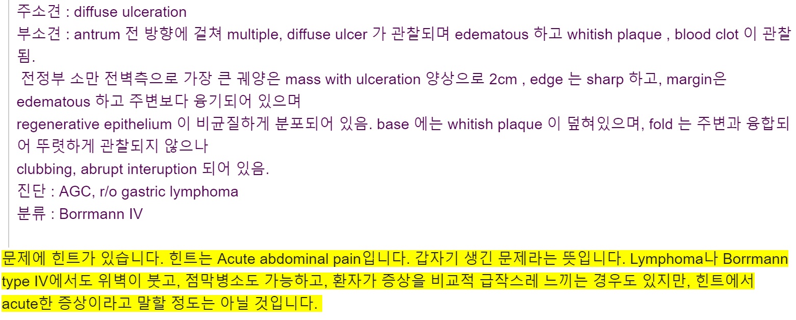
AGML ИТНРДЯДй. ДйИИ 'hemorrhageАЁ РжОњДј АЭРИЗЮ КИРг'РК ЛчСЗРдДЯДй. ГЛНУАцПЁМДТ КИРЬДТ АЭИИ ОВИщ ЕЫДЯДй. hemorrhage ПЉКЮДТ РгЛѓАЁАЁ ЦЧДмЧЯИщ ЕЩ ЙЎСІРдДЯДй. ГЛНУАц РЧЛчДТ БђВћЧЯАд КИРЬДТ АЭИИ ОЙНУДй. 'HemorrhageАЁ РжОњДј АЭРИЗЮ КИРг'РЛ ВР ОВАэ НЭДйИщ МвАпРЬГЊ impressionПЁ ОВСі ИЛАэ Бз ОЦЗЁПЁ comment (ШЄРК note)ЖѓДТ ЧзИёРЛ ИИЕщОю ЧиДч ГЛПыРЛ О№БоЧи СнНУДй.

* ТќАэ: EndoTODAY AGML
![]() Case 11
Case 11
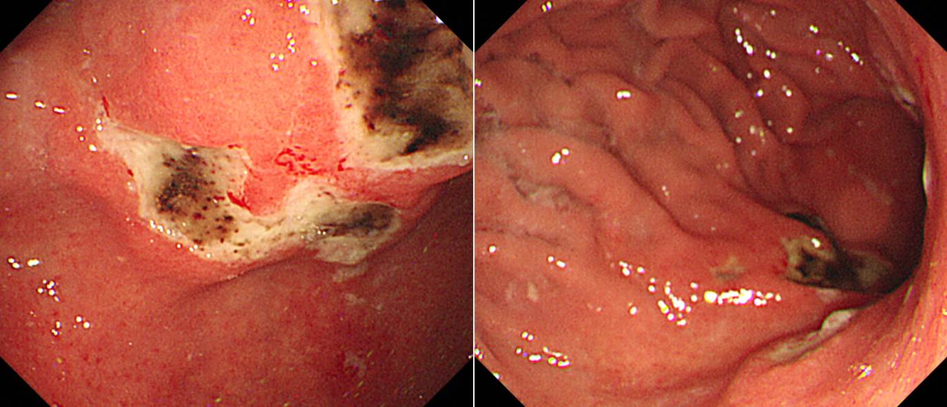
МвАп: БйРЇРќСЄКЮПЭ РЇУМЧЯКЮПЁ 2-3cm ХЉБтРЧ ДйОчЧб ХЉБтРЧ ulcerАЁ РжАэ РЯКЮ hematinРЬ КЮТјЕЧОю РжРН. Ulcer marginРЬ edematous ЧЯАэ erythemaАЁ ЕПЙнЕЧОю РжРН.
СјДм: Multiple benign gastric ulcers, A2 (r/o CMV gastritis, r/o NSAID induced gastritis)
[РЬСиЧр comment]
Multiple ulcers РЬЙЧЗЮ NSAIDs-associatedИІ АэЗСЧЯДТ АЭРЬ АЁРх ХИДчЧеДЯДй. R/O Zollinger-Ellison syndromeРЛ О№БоЧЯНХ КаЕЕ АшМЬДТЕЅПф... БлНъПф... АЁДЩМКРК ЖГОюСіДТ АЭ ААНРДЯДй.
AGMLЗЮ ДфЧб КаРЬ АшМЬНРДЯДй. БзЗЏГЊ AGMLРК КИДй acuteЧЯАэ КИДй БЄЙќРЇЧб АцПьПЁ КйРЬИщ ССАкАэ РЬ АцПьДТ ДйЙпМК БЫОчРЬ РћДчЧв АЭ ААНРДЯДй.
РЬ СѕЗЪДТ NSAID КЙПыРкРЧ ДйЙпМК БЫОчРЬОњНРДЯДй.
* ТќАэ: EndoTODAY NSAID РЇРхАќ ЧеКДСѕ
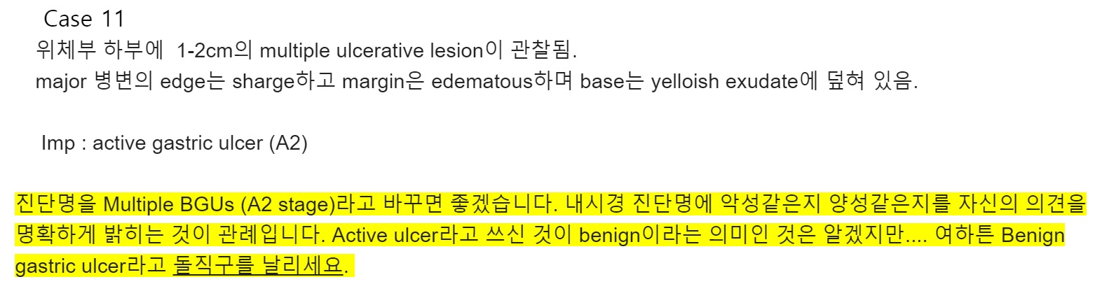
![]() Case 12
Case 12
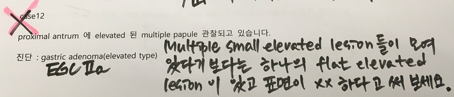
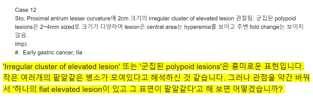
МвАп: Proximal antrum, lesser curvature ПЁ 2 cm СЄЕЕРЧ РќУМРћРИЗЮ flatЧб elevated lesionРЬ РжНРДЯДй. ЧЅИщРК nodularЧЯАэ СЄЛѓ СЁИЗАњРЧ АцАшДТ white light endoscopyПЁМДТ ДйМв КвИэШЎЧЯСіИИ indigo carmine ЛьЦї ШФПЁДТ КёБГРћ ИэШЎЧЯАд АќТћЕЫДЯДй.
СјДм: Early gastric cancer, IIa
[РЬСиЧр comment]
РќСЄКЮ РЇФЁИІ ММКаЧЯДТ ЙцЙ§РЛ ЙЎРЧЧЯНХ КаРЬ АшМЬНРДЯДй. ССРК СњЙЎРЬЖѓАэ Л§АЂЕЧОњАэ ДфКЏ ГЛПыРЛ МвАГЧеДЯДй.
СЖБтРЇОЯРЬГЊ СјЧрРЇОЯРЧ subtype КаЗљДТ ДыА ЧЯДТ АЭРЬ ССНРДЯДй (EndoTODAY СЖБтРЇОЯ ГЛНУАц КаЗљ). ДыА ГГРлЧЯЕЧ ОрАЃ ПУЖѓПдРИИщ type IIa, ЛѓДчШї ЦЂОю ПУЖѓПдРИИщ type IРИЗЮ СжНУБт ЙйЖјДЯДй. РкЗЮ УјСЄЧЯПЉ Ию mm РЬЛѓРЬИщ ОюЖЛАэ... ЕюРК ОВРпЕЅБт ОјДТ РЯРдДЯДй. СЖБтРЇОЯРЛ КАЗЮ КЛ РћРЬ ОјДТ МОчЛчЖїЕщРЛ АЁИЃФЁЗСАэ ОяСіЗЮ ИИЕч Paris КаЗљДТ ОрАЃ ЙЋНУЧиЕЕ ССНРДЯДй.

MassЗЮ ЧЅНУЧб КаРЬ АшНУДТЕЅ... РЬКИДйДТ nodular elevated lesionРИЗЮ ЧЅЧіЧиСжНУБт ЙйЖјДЯДй. Ся superficial lesionРЬЖѓДТ ЖцРЬСіПф. ГГРлЧб КДМв. MassДТ КИХы AGCПЁМ ОВДТ ПыОюРдДЯДй.
УЪНЩРк СпПЁДТ ЧдИєЧќ РЇОЯРИЗЮ КИНХ КаЕЕ АшМЬНРДЯДйИИ, РЬ КДМвДТ СжЗЮ flat nodular elevated lesionРдДЯДй. Elevated nodule ЛчРЬ ЛчРЬАЁ ОрАЃ ЧдИєЕШ АЭ ЛгРЬСі ЧдИєЧќ РЇОЯРК ОЦДеДЯДй.
ХЉБт УјСЄПЁ ДыЧб СњЙЎРЬ РжОњНРДЯДй. AntrumРЛ АЁЕц УЄПьДТ СіИЇ 6cm АјРЬ РжДйАэ Л§АЂЧи КИММПф.
РќУМИІ ЧЯГЊЗЮ КИДТ ЙцЧтРИЗЮ describe Чи КОНУДй.
ESDИІ ЧпНРДЯДй. УжСО КДИЎ АсАњДТ ОЦЗЁПЭ ААОвНРДЯДй.
Stomach, endoscopic submucosal dissection:
. Early gastric carcinoma
1. Location : antrum, lesser curvature
2. Gross type : EGC type IIa+IIb
3. Histologic type : tubular adenocarcinoma, well differentiated
4. Histologic type by Lauren : intestinal
5. Size : 2.8x2.3 cm
6. Depth of invasion : invades mucosa (lamina propria) (pT1a)
7. Resection margin: free from carcinoma (safety margin: distal 0.8 cm, proximal 0.8 cm, anterior 0.4 cm, posterior 0.8 cm)
8. Lymphatic invasion : not identified
9. Venous invasion : not identified
10. Perineural invasion : not identified
11. Microscopic ulcer : absent
12. Histologic heterogeneity: absent
2017Гт МвШБтЧаШИ ПЌМіАСТПЁМ РЇОЯРЧ СјДмАњ ФЁЗсПЁ ДыЧЯПЉ АРЧЧб ОЦЗЁ ЕППЕЛѓРЛ ТќАэЧи СжНУБт ЙйЖјДЯДй.
![]() СѕЗЪ 13.
СѕЗЪ 13.
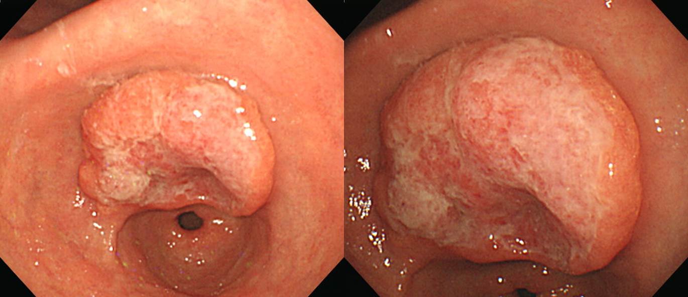
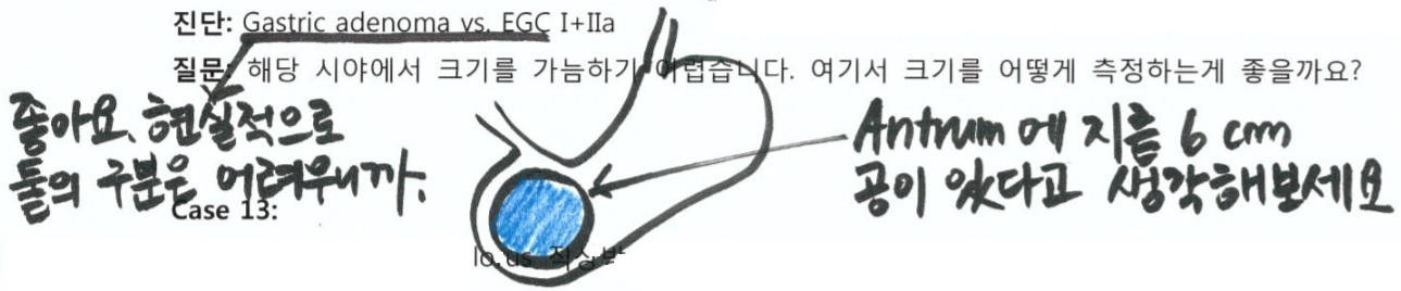
МвАп: РЇРќСЄКЮ МвИИПЁ 4 cm ХЉБтРЧ massАЁ РжРН. MassРЧ ЛѓДмКЮДТ ЧдИєЕЧОю РжДТЕЅ distal КЮРЇДТ СжКЏПЁ КёЧЯПЉ РЯКЮ ЧдИєЕЧОю РжРН.
СјДм: AGC, type I
[РЬСиЧр comment]
EGCРЮСі AGCРЮСі ОжИХЧеДЯДй. ДйМв БэРК EGCРЯ МіЕЕ РжАэ, PM cancer СЄЕЕРЧ AGCРЯ МіЕЕ РжДТ БзЗБ ЛѓШВРЮЕЅПф.... МіМњ ШФ КДИЎАсАњПЁМДТ deep SM invasion (SM3)РЛ КИРЮ EGCПДСіИИ, ГЛНУАц СјДмРИЗЮ AGCИІ КйПДДйАэ ХЉАд ХПЧв СЄЕЕДТ ОЦДеДЯДй. EGC type IАњ AGC type IРК ДУ БИКаЧЯБтАЁ ОюЗЦБт ЖЇЙЎРдДЯДй (EndoTODAY КИИИ 1Чќ СјЧрМК РЇОЯАњ 1Чќ СЖБтРЇОЯРЧ БИКа).
AGCЗЮ КИОвРЛ ЖЇ КИИИ ХИРдРЬ ДйМв ОжИХЧеДЯДй. РњДТ КИИИ 1ЧќРЛ УпУЕЧеДЯДй. РЯКЮАЁ ДйМв ЧдИєЕЧОю РжСіИИ КИИИ 2ЧќПЁМ КИРЬДТ ЖбЗЧЧЯАэ БэРК БЫОчРК ОЦДЯДЯБюПф.
7-8НУ ЙцЧтРЧ abnormal foldПЁ ДыЧб СњЙЎРЬ РжОю ДфЧеДЯДй. РЯЙнРћРИЗЮ foldДТ БЫОчЧќ КДМвПЁМ ЙпЛ§ЧЯСіИИ РЖБтЧќ КДМвПЁМЕЕ СЁИЗРЬ ЕщИЎИщМ СжИЇРЬ РтШїДТ АцПьАЁ РжНРДЯДй. SMTПЁМДТ РЬЗБ АЭРЬ РкСж КИРЬДТЕЅ bridging foldЖѓАэ ЧеДЯДй. РЬПЭ КёНСЧЯДйАэ РЬЧиЧЯНУБт ЙйЖјДЯДй. ДйИЅ ЦЏКАЧб commentДТ ОјНРДЯДй. РЬСиЧр
AGCЗЮ Л§АЂЧЯАэ МіМњЧпДТЕЅ deep SM invasionРЛ КИРЬДТ EGCПДНРДЯДй.
Stomach, subtotal gastrectomy:
Early gastric carcinoma
1. Location : lower third, Center at antrum and lesser curvature
2. Gross type : EGC type I
3. Histologic type : gastric carcinoma with lymphoid stroma (medullary carcinoma)
4. Histologic type by Lauren : intestinal
5. Size : 3x2.3 cm
6. Depth of invasion : invades submucosa (sm3) (pT1b)
7. Resection margin: free from carcinoma, safety margin: proximal 17 cm, distal 4.5 cm
8. Lymph node metastasis : no metastasis in 41 regional lymph nodes (pN0), (perinodal extension: absent)
9. Lymphatic invasion : present
10. Venous invasion : not identified
11. Perineural invasion : not identified
12. Peritoneal cytology : negative
13. AJCC stage by 7th edition: T1b N0
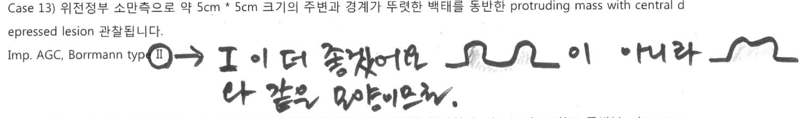
2017Гт ЛѕЗЮ fellow АњСЄПЁ ЕщОюПТ МБЛ§ДдВВМ АњАХ КЛРЮРЬ АцЧшЧб РЏЛч СѕЗЪИІ КИГЛСжМЬНРДЯДй. КДИЎ typeРЬ ЕПРЯЧб АЭРЬОњНРДЯДй. АЈЛчЧеДЯДй.
Stomach, total gastrectomy ;
EARLY GASTRIC CARCINOMA
Location : upper third
Gross type : EGC type IIa
Histologic type: MODERATELY DIFFERENTIATED ADENOCARCINOMA WITH LYMPHOID STROMA (=lymphoepithelioma-like carcinoma)
Histologic type by Lauren: Intestinal type
Size: 1.7 x 1.2 cm
Depth of invasion: invades submucosa (sm3) (pT1b)
Resection margin: free from carcinoma
NO PERIGASTRIC LYMPH NODE available for histologic examination
Lymphatic invasion: not identified
Venous invasion: not identified
Perineural invasion: not identified
Pathologic stage: pT1b pNX
ГЛФЃ БшПЁ lymphoepithelioma-like carcinoma (ИВЧСОчЛѓЧЧСООч ОЯСО)ИІ СЖБн Дѕ МГИэЧи КИАкНРДЯДй. Lymphoepithelioma-like carcinoma (LELC)ДТ КёРЮЕЮОЯАњ СЖСїЧаРћРИЗЮ РЏЛчЧЯАэ, ИВЧСБИМК АЃСњРЛ АЁСј ЙЬКаШЕШ РЇСООчРдДЯДй. Epstein-Barr ЙйРЬЗЏНКПЭРЧ ПЌАќМКЕЕ О№БоЕЧАэ РжНРДЯДй. LELCРК НФЕЕ, РЇ, ДуАќ, ХИОз МБ, Цѓ, РкБУ, ЧЧКЮ ЕюПЁМ ЙпЛ§ЧеДЯДй. РЇ LELCДТ РќУМ РЇОЯРЧ l-4%ИІ ТїСіЧЯДТЕЅ СЖБтРЇОЯРЧ АцПь IIa + IIcЧќРЬ ИЙРИИч СјЧрМК РЇОЯРЧ АцПь Borrmann type IV ЧќРЬ ИЙДйАэ ЧеДЯДй. СЖБтРЇОЯРЧ ЧќХТЗЮ ГЊХИГЏ АцПьДТ РЯЙнРћРЮ СЖБтРЇОЯАњ ПЙШФАЁ КёНСЧЯГЊ СјЧрМК РЇОЯРЧ ЧќХТИІ УыЧв АцПь РЯЙнРћРЮ СјЧрМК РЇОЯПЁ КёЧЯПЉ ПЙШФАЁ ШЮОР ССРК АЭРИЗЮ ОЫЗССЎ РжНРДЯДй. ЕхЙААд СЁИЗЧЯ СООчРЧ ЧќХТЗЮ ГЊХИГЊДТ АцПьЕЕ РжОю СжРЧАЁ ЧЪПфЧеДЯДй. (ТќАэ: ДыЧбМвШБтГЛНУАцЧаШИСі 2004;28:123-126)
АќЗУ РњГЮ ЧЯГЊ МвАГЧеДЯДй.
Wang HH, Wu MS, Shun CT, Wang HP, Lin CC, Lin JT. Lymphoepithelioma-like carcinoma of the stomach: a subset of gastric carcinoma with distinct clinicopathological features and high prevalence of Epstein-Barr virus infection. Hepatogastroenterology. 1999;46(26):1214-9.
METHODOLOGY: Of 379 patients with gastric adenocarcinoma, from 1993 to 1996, 6 of them with lymphoepithelioma-like carcinoma of stomach were retrospectively studied.
RESULTS: Five patients were females and one patient was male. Their age ranged from 51-75 years with a mean age of 61.5 years. Endoscopically, 2 patients were initially diagnosed as early gastric cancer and the other 4 were diagnosed as advanced gastric cancer. Three patients had tumors located in the lower third of the stomach, while the other three tumors were located in the middle and upper third. Two tumors invaded into the serosal layer and the other four lesions were confined at submucosal and muscular layers. Using the in situ hybridization method, all 6 patients (100%) had positive nuclear Epstein-Barr virus-encoded small RNA signals in the tumor cells but not in the surrounding lymphoid stroma and non-neoplastic gastric mucosa. Helicobacter pylori was found in 4 (66.7%) of the cases. The mean follow-up period of the 6 patients was 27 months. Five patients were free of the disease. Lymph node involvement and mesenteric implantation was noted in one patient in which cancer recurred 1 year after gastrectomy.
CONCLUSIONS: Lymphoepithelioma-like carcinoma of stomach in this study revealed a female predominance, preferential localization in the proximal part of the stomach, better prognosis, and a high association with Epstein-Barr virus infection.
АќЗУ СѕЗЪ Ию АГ МвАГЧеДЯДй.
Early gastric carcinoma
1. Location : lower third, center at antrum and greater curvature
2. Gross type : EGC type IIc+IIa
3. Histologic type : lymphoepithelioma-like carcinoma (EBV-positive)
4. Histologic type by Lauren : intestinal
5. Size : 2.5x2x0.5 cm
6. Depth of invasion : extension to submucosa (sm3) (pT1b)
7. Resection margin: free from carcinoma, safety margin: proximal, 10.0 cm; distal, 1.8 cm
8. Lymph node metastasis : metastasis to 2 out of 36 regional lymph nodes (pN1)
9. Lymphatic invasion : not identified
10. Venous invasion : not identified
11. Perineural invasion : not identified
Advanced gastric carcinoma
1. Location : upper third, Center at high body and greater curvature
2. Gross type : Borrmann type 2
3. Histologic type : lymphoepithelioma-like carcinoma
4. Histologic type by Lauren : diffuse
5. Size : 4.4x3.2x1.4 cm
6. Depth of invasion : extension to proper muscle (pT2a)
7. Resection margin: free from carcinoma, safety margin: proximal, 5.5 cm; distal, 21 cm
8. Lymph node metastasis : no metastasis in 67 regional lymph nodes (pN0)
9. Lymphatic invasion : not identified
10.Venous invasion : not identified
11.Perineural invasion : not identified

1. Location : upper third, center at cardia (Siewert III)
2. Gross type : Borrmann type 2
3. Histologic type : Medullary carcinoma (lymphoepithelioma-like carcinoma)
4. Histologic type by Lauren : indeterminate
5. Size : 2.2x2.2 cm
6. Depth of invasion : invades muscularis propria
7. Resection margin: free from carcinoma, safety margin: proximal 1.4 cm, distal 2 cm
8. Lymph node metastasis : no metastasis in 52 regional lymph nodes
9. Lymphatic invasion : not identified
10. Venous invasion : not identified
11. Perineural invasion : not identified
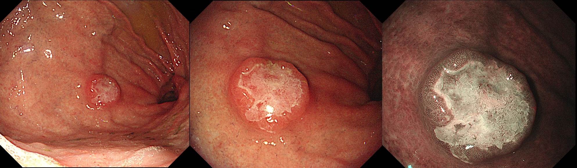
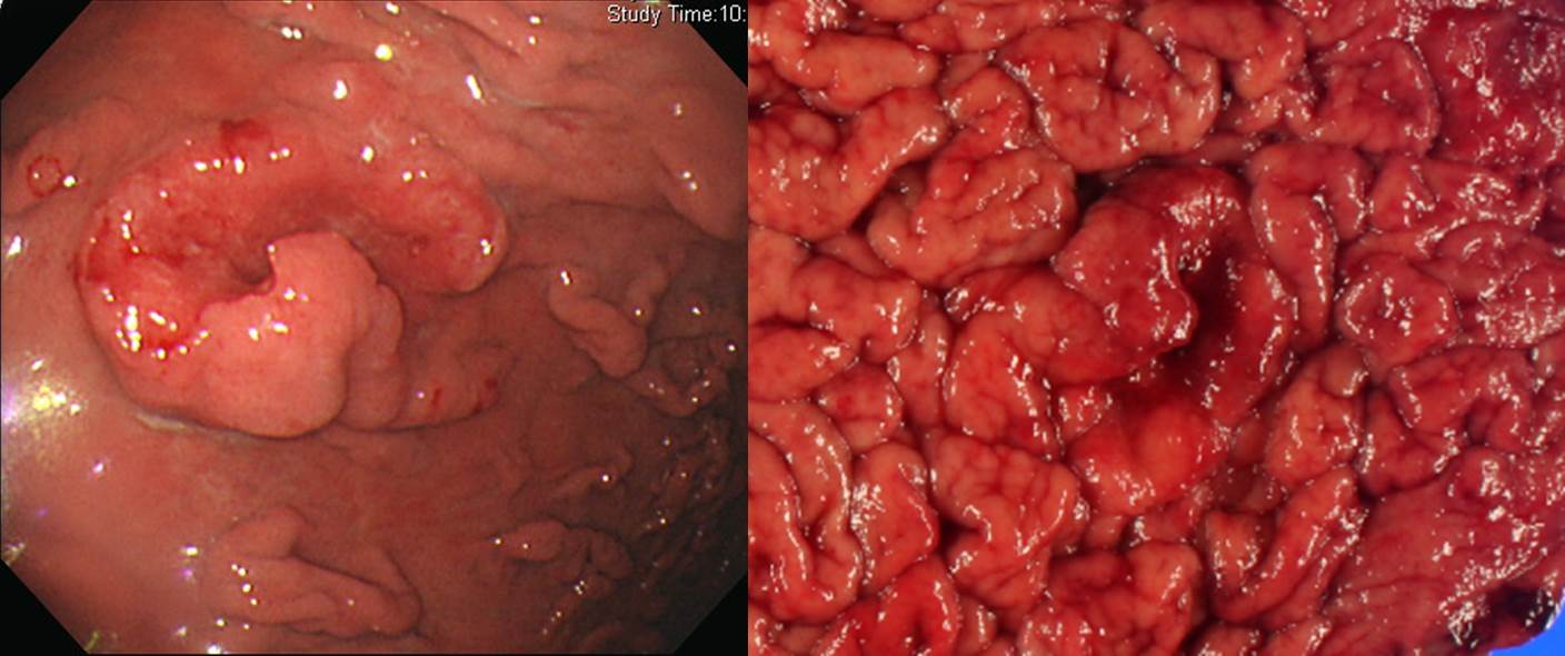
![]() Case 14. Duodenal bulb.
Case 14. Duodenal bulb.
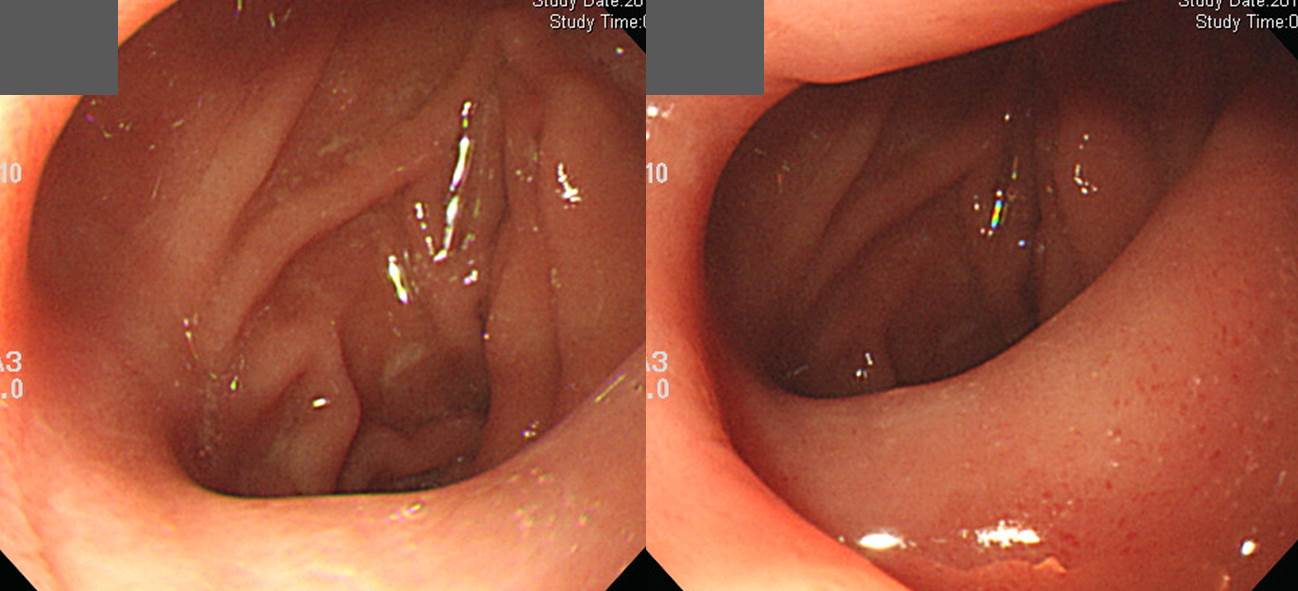
Findings: НЪРЬСіРх БИКЮРЧ РќКЎТЪРИЗЮ foldЕщРЬ convergingЧЯДТ И№ОчРЬАэ ulcer craterДТ ОјРН. РЬЗЮ РЮЧЯПЉ НЪРЬСіРх БИКЮАЁ ОрАЃ deformed ЕЧОю РжРН. (Duodenal bulb was deformed due to multiple converging folds. There was no active ulcer crater.)
Impression: Duodenal ulcer scar, S2
[РЬСиЧр comment]
НЪРЬСіРхРЬ ИХПь ОюЗСПќДј И№ОчРдДЯДй. РлРК erosion ШЄРК ulcerative lesionРЬЖѓЕЕ ДфЧб КаЕЕ АшНУДТЕЅПф... ЛчНЧ БзЗЏЧб СОЗљДТ ОЦЙЋ АЭЕЕ ОјНРДЯДй. ДмСі ulcer scarПЭ СжКЏРИЗЮ ВјЗСПРДТ ПЉЗЏ fold ЕщИИ РжРЛ ЛгРдДЯДй.
КИРЬСі ОЪДТ ulcer craterИІ КИРЮДйАэ ЧЯАэ, КИРЬДТ foldИІ КИРЬСі ОЪДТДйАэ ДфЧб КаЕщРЬ ИЙНРДЯДй. МБРдАпРЬ РЬИЎ ЙЋМЗНРДЯДй. ОЦЗЁПЭ ААРЬ ЧиМЎЧЯБт ЙйЖјДЯДй.
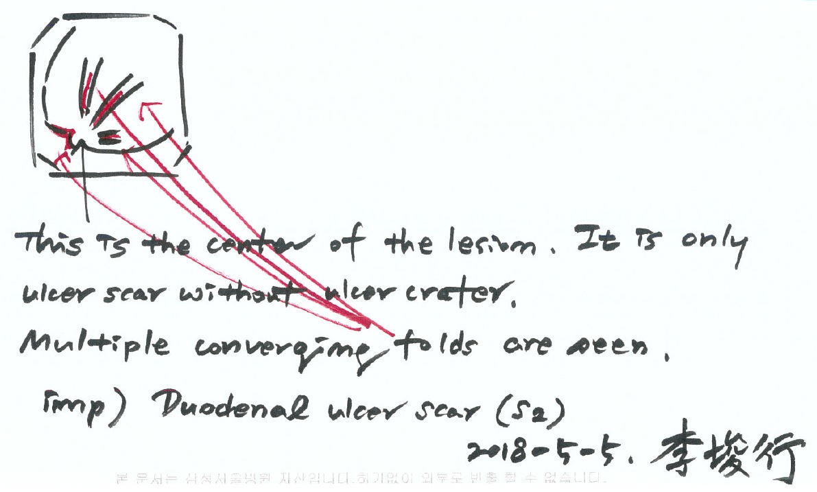
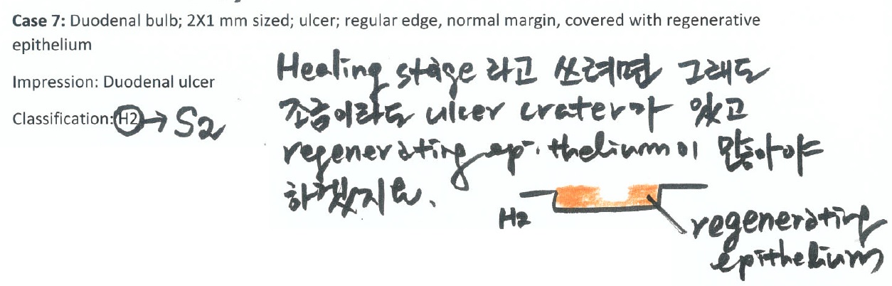
H2 stageЗЮ ДфЧиСжНХ КаРЬ АшМХМ ОЦЗЁПЭ ААРЬ БзИВРЛ БзЗС МГИэЧЯПДНРДЯДй.
ОЦЗЁДТ melena ИчФЅ ШФ ГЛПјЧЯМЬДј КаРдДЯДй. УтЧїРК ОјОњАэ ulcer craterПЭ red pigmentation СЄЕЕИИ КИПДНРДЯДй. УпРћГЛНУАцПЁМ ПЯРќШї ШЃРќЕЧОњНРДЯДй. ИИОр ОюЖВ ШЏРкПЁМ СТУј ЛчСјАњ ААРК МвАпРЬ КИРЬИщ БЫОчРЧ АњАХЗТРЛ ЙАОюКИПЉОп ЧЯАкСіПф?
![]() [EndoTODAY Weekly Seminar ДйНУКИБт (2020Гт)]
[EndoTODAY Weekly Seminar ДйНУКИБт (2020Гт)]
2020-25. СпШЏРкНЧ ГЛНУАцАњ НКЦЎЗЙНК БЫОч ПЙОр

2020-24. КИИИ 4Чќ СјЧрМКРЇОЯ. AGC Borrmann type 4
2020-23. Achalasia, lymphoma, EG junction cancer, Trichuris trichiura, EGC arising from hyperplastic polyp
2020-22. Esophageal diverticulum, anal melanoma, Henoch Scholein purpura, colonic invasive aspergillosis, esophageal neuroendocrine carcinoma
2020-21. Hiatal hernia Achalasia EGC Cowden NET (ДыЧбРЇДыРхГЛНУАцЧаШИ on-line АРЧ) - ЧаШИ 1До ШФРЮ 7Пљ 7РЯКЮХЭ YouTubeПЁМ НУУЛЧЯНЧ Мі РжНРДЯДй.
2020-20. Cowden syndrome ФЋПьЕЇ СѕШФБК
2020-19. Inlet patch, lymphangioma, cavernous hemanioma, rectal carcinoid
2020-18. FAP. Familial adenomatous polyposis. АЁСЗМК МБСОМК ПыСОСѕ
2020-17. Hereditary non-polyposis colorectal cancer (HNPCC) = Lynch syndrome, РЏРќМК ДыРхОЯ
2020-16. ЙйЗП НФЕЕОЯ, РЇНХАцГЛКаКёСООч, ЧуЧїМК РхПА, ДыРх РќРЬ
2020-15. ЙЬЖѕРЮАЁ, БЫОчРЮАЁ, ШЄНУ ОЯРК ОЦДбАЁ?
[Special] ЧяИЎФкЙкХЭПЁ ДыЧб ЛчАп (2020)
2020-14. РЇОЯ, РЇПыСОСѕ (Cowden syndrome), ГЛНУАц ЛчСјАњ ГЛНУАц НУНКХлРЧ button РЭШїБт
2020-13. НФЕЕОЯ, amyloidosis, extrinsic compression
[Special] МјУЕИИ ГЛНУАц ММЙЬГЊ ЦЏА: РЇРхАќ АдНЧСѕ
2020-12. Introduction to IEE (image enhanced endoscopy)
2020-11. One point lesson - РРБоГЛНУАцАњ БнНФБтАЃ, actinomycosis, CMV НФЕЕПА
2020-10. НФЕЕСњШЏ ГЛНУАц ЛчСј ХфЗа. Mucosal break, hiatal hernia, achalasia, esophageal ESD
2020-9. Quiz ЧиМГ (МКАс) - ЛѓКЮНФЕЕОЯ, РЇОЯАњ РЇБЫОч, ЧзЙЎ ШцЛіСО, СїРх MALToma
2020-8. РЇУрМК РЇПА, ПЭЦФИА, ГФЁМК БЫОч, КЏКё
2020-7. Quiz ЧиМГ (ПРСжЧі)
2020-6. Quiz ЧиМГ (БшЙЮСі)
2020-5. НФЕЕ СњШЏ FAQ. ЙЋСѕЛѓ НФЕЕ ФЕ№ДйСѕ, Sloughing esophagitis, НФЕЕ black pigmentation, НФЕЕ dysplasia, Achalasia, Diffuse idiopathic skeletal hyperostosis
2020-4. ГЛНУАц АЫЛч Рќ ОЦНКЧЧИА СпДм, ЧяИЎФкЙкХЭ СЖСїАЫЛч, MALToma УпРћАЫЛч Ею fellow МБЛ§ДдЕщРЧ СњЙЎПЁ ДфЧеДЯДй.
2020-3.
2020-2.
2020-1.
© РЯПјГЛНУАцБГНЧ ЙйИЅГЛНУАцПЌБИМв РЬСиЧр. EndoTODAY Endoscopy Learning Center. Lee Jun Haeng.