 EndoTODAY 내시경 교실
Beginner | ESA | Schedule | OPD
Seminars | Atlas | Recent | Links
![]() [일원내시경교실 목요점심집담회 2016-5-12]
[일원내시경교실 목요점심집담회 2016-5-12]
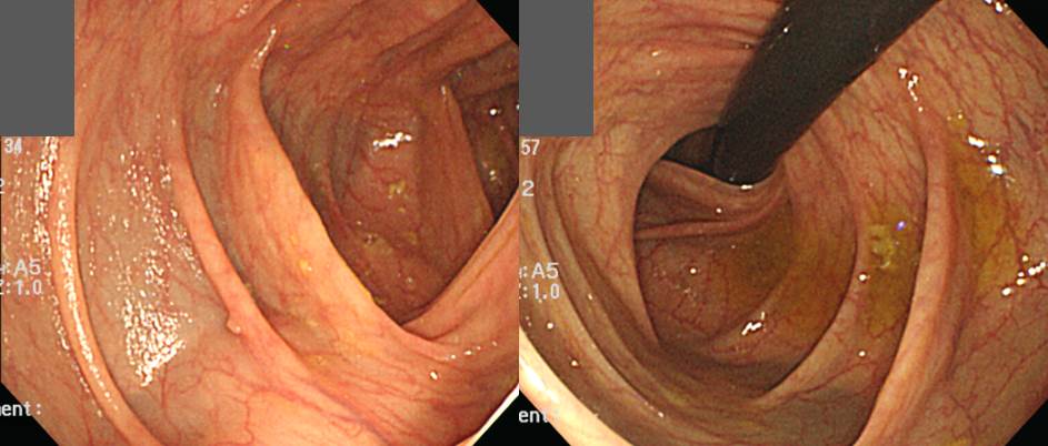
![]() 1. Hiatal hernia, sliding type
1. Hiatal hernia, sliding type
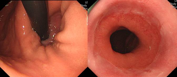
기준이 2cm에 가까스로 해당하는 정도의 경미한 hiatal hernia로 보면 좋겠습니다.
* 참고: EndoTODAY Hiatal hernia
![]() 2. Borrmann type IV 첫 증례
2. Borrmann type IV 첫 증례
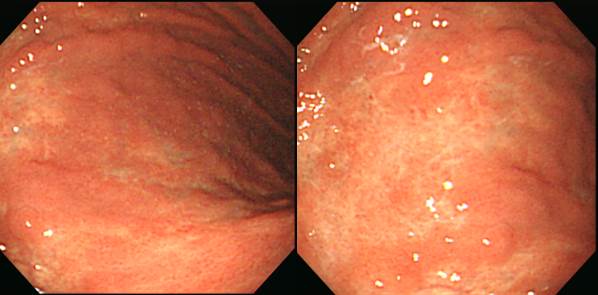
보만 4형 진행성 위암은 정말 놓치기 쉽습니다. 이런 형태는 더욱 그러합니다. 표면이 위축성 위염처럼 보이는 경우였는데 조직검사에서 P/D adenocarcinoma가 나왔습니다.
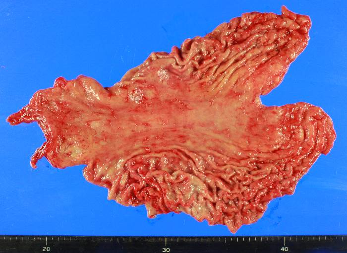
Stomach, total gastrectomy::
Advanced gastric carcinoma
1. Location : upper third, Center at body and greater curvature
2. Gross type : Borrmann type 4
3. Histologic type : tubular adenocarcinoma, poorly differentiated
4. Histologic type by Lauren : diffuse
5. Size : 12.2x8.3 cm
6. Depth of invasion : penetrates subserosal connective tissue (pT3)
7. Resection margin: free from carcinoma, safety margin: proximal 0.9 cm, distal 15.9 cm
8. Lymph node metastasis : no metastasis in 81 regional lymph nodes (pN0)
9. Lymphatic invasion : not identified
10. Venous invasion : not identified
11. Perineural invasion : present
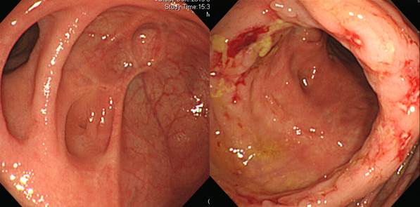
![]() 3. Borrmann type IV 두번째 증례
3. Borrmann type IV 두번째 증례
Stomach, total gastrectomy:
Advanced gastric carcinoma
1. Location : upper third, Center at body and posterior wall
2. Gross type : Borrmann type 3
3. Histologic type : tubular adenocarcinoma, poorly differentiated
4. Histologic type by Lauren : diffuse
5. Size : 4x3 cm
6. Depth of invasion : invades serosa (pT4a)
7. Resection margin: free from carcinoma, safety margin: proximal 2 cm, distal 7.5 cm
8. Lymph node metastasis : metastasis to 6 out of 45 regional lymph nodes (pN_)
9. Lymphatic invasion : present
10. Venous invasion : not identified
11. Perineural invasion : present
12. Peritoneal cytology : negative
아래는 집담회 증례를 준비하신 임상강사 선생님께서 정리한 보만 4형 진행성 위암의 특징입니다.
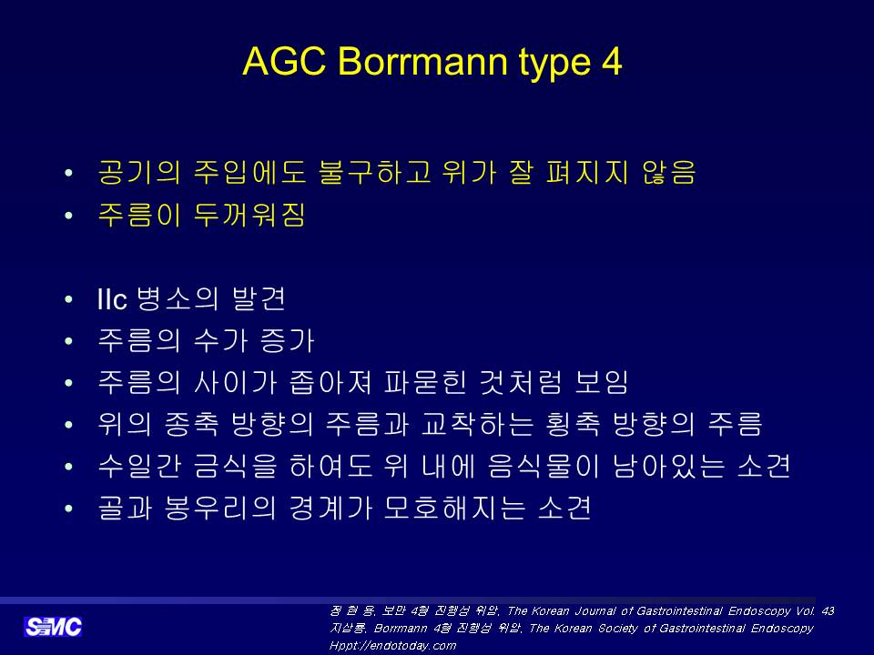
* 참고: Borrmann type IV 연제: EndoTODAY 20130211 부터 EndoTODAY 20130306
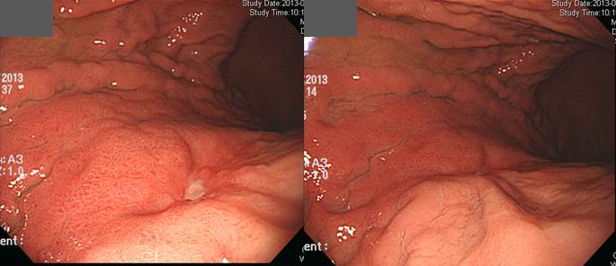
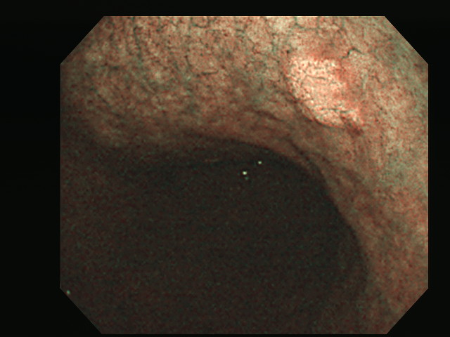
![]() 4. 결핵성 장염
4. 결핵성 장염
Cecum에서는 scar change가 있었고 다른 부위에는 active 장염이 있었던 경우입니다.
아래는 집담회 증례를 준비하신 임상강사 선생님께서 정리한 결핵성 장염의 특징입니다.
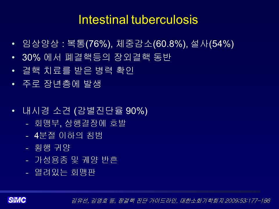
* 참고: EndoTODAY 결핵성 위장질환
![]() 5. 어떤 대장암 (원발성 혹은 전이성)
5. 어떤 대장암 (원발성 혹은 전이성)
우연히 시행한 CA-19-9가 400U/ml 정도로 높아서 PET-CT 후 소화기내과를 방문한 환자입니다.
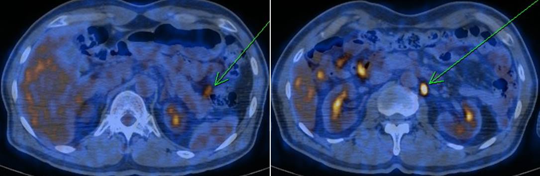
PET에서 "Left paraaortic lymph node, left mesenteric lymph node에서 SUVmax = 6.6으로 측정되는 국소 hypermetabolic lesion이 관찰되는데 malignancy의 metastasis 가능성이 높겠음. Hepatic flexure 부위 colon에서 SUVmax = 5.6으로 측정되는 hypermetabolic lesion이 관찰되는데 primary malignancy의 가능성이 있어 colonoscopy를 권장함" 소견이었고 대장내시경을 하였는데 대장내시경에서 작은 용종 이외에는 특이소견이 없었습니다.
확인을 위하여 explore laparotomy를 시행하였습니다. 수술 소견은 "splenic flexure 주변의 mesocolon 에 poorly demarcated hard mass 있어, Lt.colon을 mobilization 한 후에 Lt.colic artery를 따라 hard LNs있어 dissection 시행 후 en bloc 으로 colon을 segmental resection and anastomosis 시행함, S2 에 2cm sized mass 있어 margin 확보하기 위해 Lt.lateral segmentectomy 시행함"으로 되어 있었고 최종 병리는 아래와 같았습니다.
Colon, segmental resection:
Adenocarcinoma, moderately differentiated
1. Location: splenic flexure of colon
2. Gross type: unclassifiable
3. Size: 3x2x1.5 cm
4. Depth of invasion: 1) mainly involved pericolic adipose tissue without involvement of mucosa, 2) penetrates visceral peritoneum(pT4a)
5. Resection margin: free from carcinoma, safety margin: nearest, 2.5 cm ; opposite, 3 cm ; radial, > 5mm
6. Regional lymph node metastasis : metastasis to 11 out of 12 regional lymph nodes(pN2b)
7. Lymphatic invasion: present
8. Venous invasion: present (extramural)
9. Perineural invasion: present (++)
10. Tumor budding : negative
11. Associated findings : metastasis
12. Pathologic staging: pT4a N2b M1a (liver)
병리과에서는 대장암처럼 결과를 주셨지만 저는 (1) 원발부위가 대장암이고 간전이가 동반된 것인지, (2) 어디선가 unknown origin의 암이 간과 대장에 전이된 것인지 확신이 들지 않았습니다. 점막층에는 암이 없고 주로 pericolic adipose tissue에만 암세포가 존재하는 대장암을 만난 적이 없기 때문입니다.
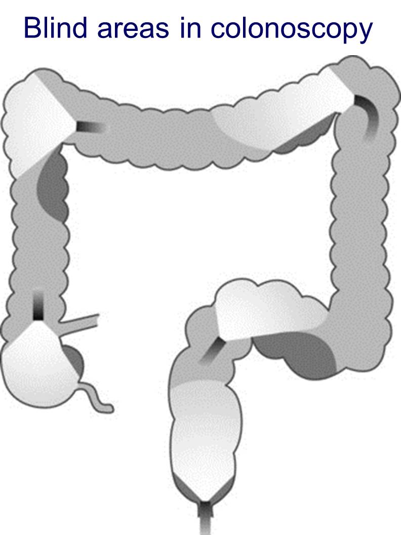
여하튼 대장암이라 가정하고 말씀드리면, 대장내시경에서는 질병이 발견되지 않았습니다. CA-19-9 상승으로 인하여 explo lapa를 통하여 대장암이 진단된 드문 경우였습니다. 대장내시경에는 항상 blind area가 있다는 것을 명심합시다.
저는 건강검진에 CA 19-9가 포함되는 것을 반대합니다. 득을 보는 사람은 별로 없고 손해보는 사람은 많은 것 같습니다 (개인적인 느낌).
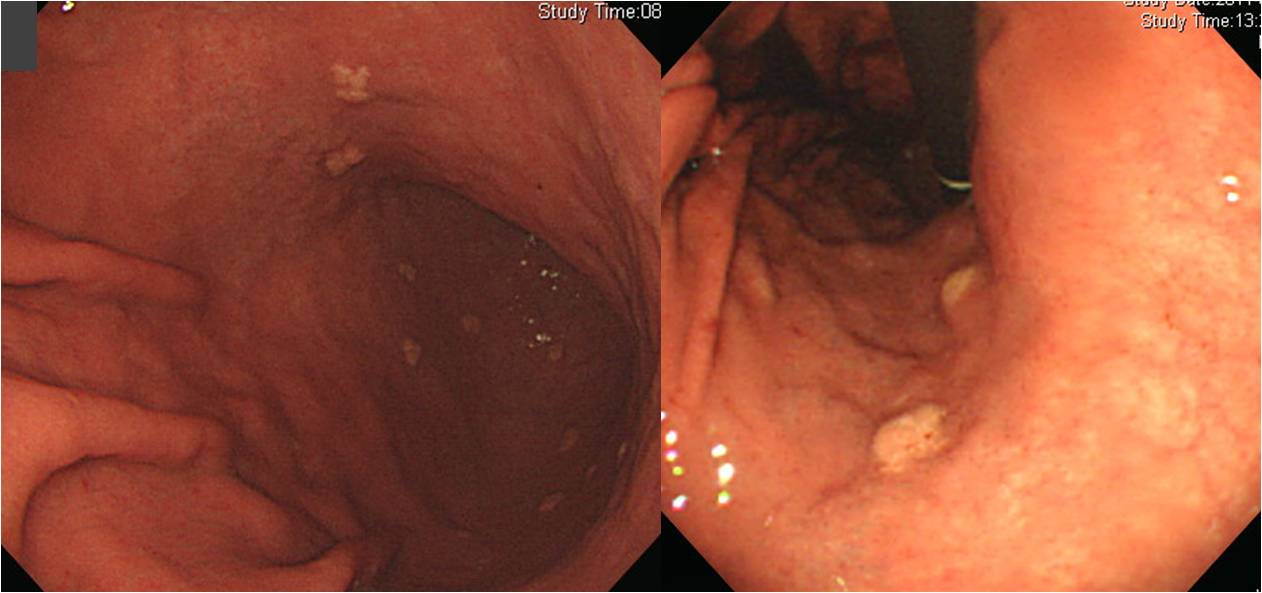
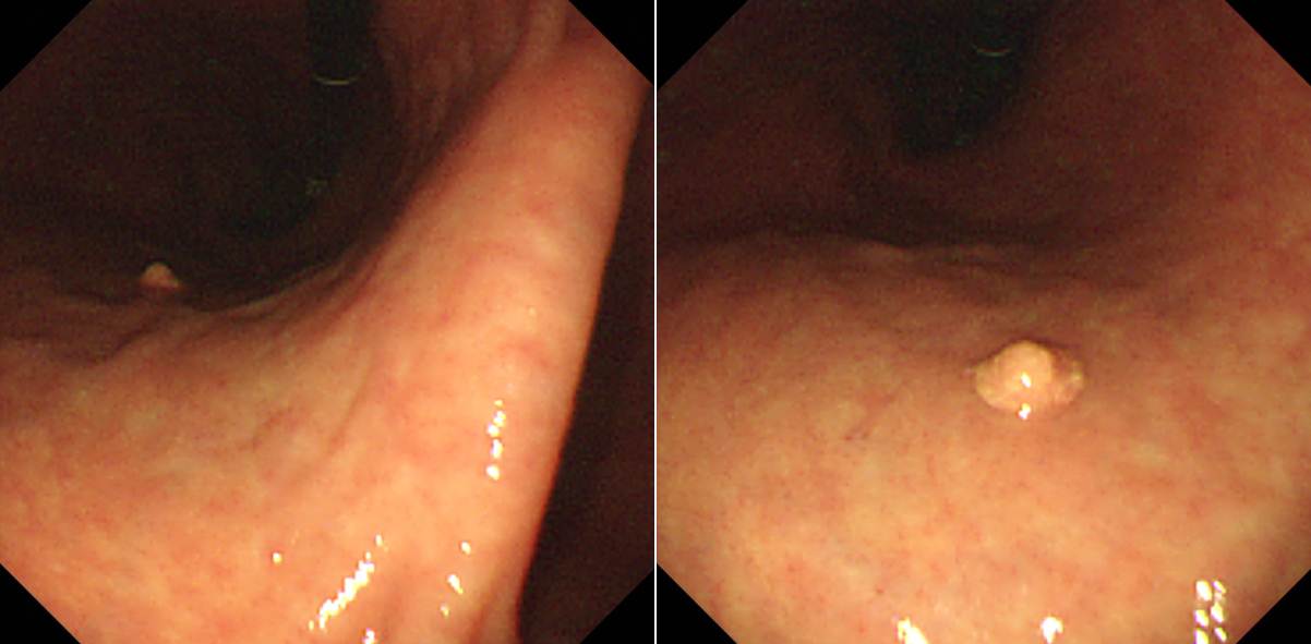
![]() 6. Xanthoma
6. Xanthoma

Xanthoma는 flat 하다고 생각하는 분이 많지만 이 증례처럼 융기된 경우도 많습니다. 다양한 xanthoma입니다.


* 참고: 최근 대한상부위장관헬리코박터 학회지에 실린 리뷰가 있어 소개합니다.
![]() [References]
[References]
1) SMC Endoscopy Unit 삼성서울병원 내시경실
2) SMC Monday GI conference 삼성서울병원 일원내시경교실 월요점심소화기집담회
3) SMC Thursday endoscopy conference 삼성서울병원 일원내시경교실 목요점심내시경집담회
© EndoTODAY Endoscopy Learning Center. Jun Haeng Lee.