EndoTODAY 내시경 교실
Beginner | ESA | Schedule | OPD
Seminars | Atlas | Recent | Links
![]() [Thursday Endoscopy Conference 20161027]
[Thursday Endoscopy Conference 20161027]
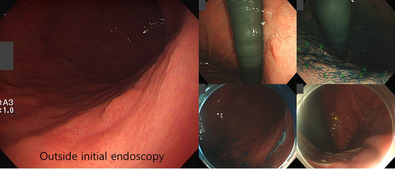
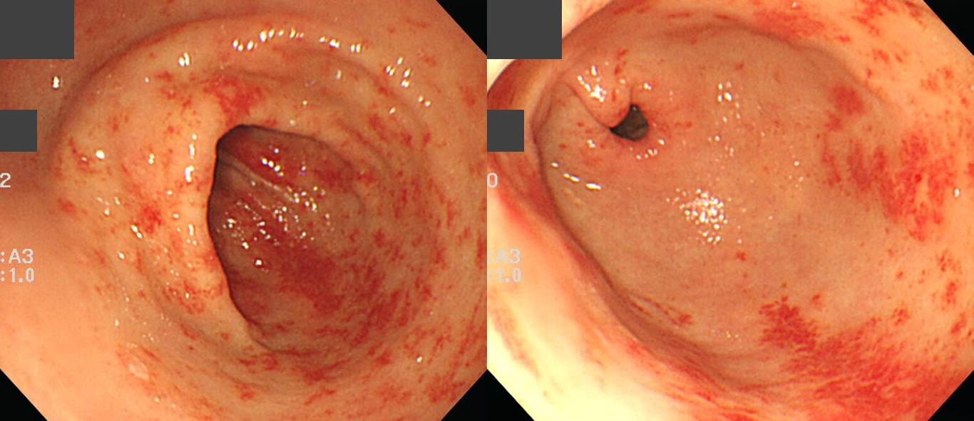
![]() 1. EGC (WHYX type)
1. EGC (WHYX type)
내시경 검사에서 15mm 크기의 EGC IIa로 판단되었습니다. ESD 후 최종 결과는 26mm WHYX 병소였습니다. 분화도가 매우 좋아 경계 판단이 어려운 종류의 위암입니다.
Stomach, endoscopic submucosal dissection:
Early gastric carcinoma
1. Location : body, posterior wall
2. Gross type : EGC type IIb
3. Histologic type : tubular adenocarcinoma, moderately differentiated (WHYX lesion)
4. Histologic type by Lauren : intestinal
5. Size of carcinoma : (1) longest diameter, 26 mm (2) vertical diameter, 17 mm
6. Depth of invasion : invades mucosa (lamina propria) (pT1a)
7. Resection margin : free from carcinoma(N), safety margin : distal 10 mm, proximal 12 mm, anterior 12 mm, posterior 16 mm
8. Lymphatic invasion : not identified(N)
9. Venous invasion : not identified(N)
10. Perineural invasion : not identified(N)
11. Microscopic ulcer : absent
12. Histologic heterogeneity: absent
* 참고: EndoTODAY 절제 변연 양성
![]() 2. GAVE (gastric antral vascular ectasia)
2. GAVE (gastric antral vascular ectasia)
70대 여성. 반복되는 melena. 몇 번의 APC ablation 후 호전되었습니다.
* 참고: EndoTODAY GAVE
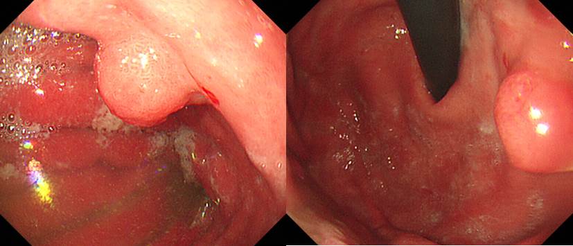
![]() 3. Remnant gastric cancer with gastritis cystica profunda
3. Remnant gastric cancer with gastritis cystica profunda
잔위암이었습니다. ESD를 시행하였습니다. 병소의 깊이에 비하여 조금 더 많이 돌출된 형태였습니다. Gastritis cystica profunda가 있었기 때문으로 추정하였습니다.
Stomach, endoscopic submucosal dissection:
Early gastric carcinoma
1. Location : high body, lesser curvature
2. Gross type : EGC type IIa
3. Histologic type : tubular adenocarcinoma, moderately differentiated
4. Histologic type by Lauren : intestinal
5. Size of carcinoma : (1) longest diameter, 12 mm (2) vertical diameter, 9 mm
6. Depth of invasion : invades mucosa (lamina propria) (pT1a)
7. Resection margin : free from carcinoma(N), safety margin : distal 2 mm, proximal 2 mm, anterior 4 mm, posterior 2 mm
8. Lymphatic invasion : not identified(N)
9. Venous invasion : not identified(N)
10. Perineural invasion : not identified(N)
11. Microscopic ulcer : absent
12. Histologic heterogeneity: absent
13. Associated finding: gastritis cystica profunda
* 참고: EndoTODAY 잔위암
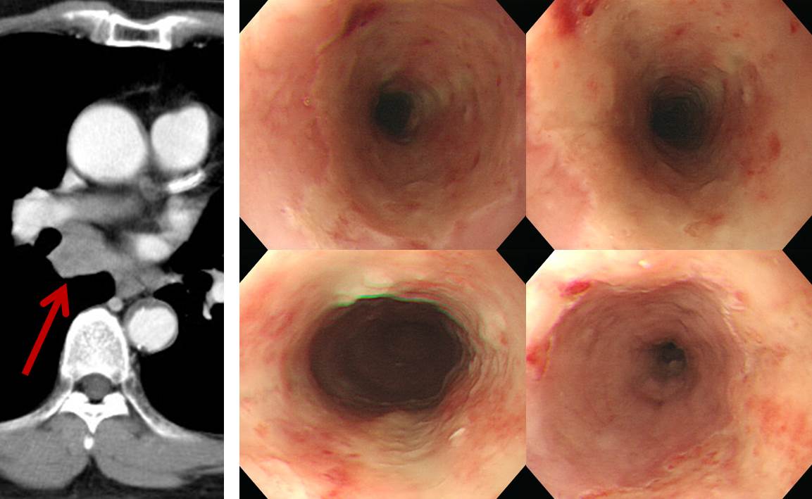
![]() 4. CMV esophagitis after CCRT for small cell lung cancer
4. CMV esophagitis after CCRT for small cell lung cancer
소세포폐암으로 definitive CCRT를 받은 환자로 neutropenia 상태에서 dysphagia가 발생하였습니다. 상절치로부터 23-30cm 병소였습니다.
RT-induced esophagitis를 생각하신 분도 계셨으나 경계가 명료하지만 약간 지도상을 보이는 점, 방사선 치료 후 전형적인 edema는 별로 없다는 점, 표면이 viral esophagitis와 유사하다는 점을 고려한다면 CMV 식도염을 먼저 생각해야 하는 증례입니다.
* 참고: EndoTODAY 바이러스 식도염
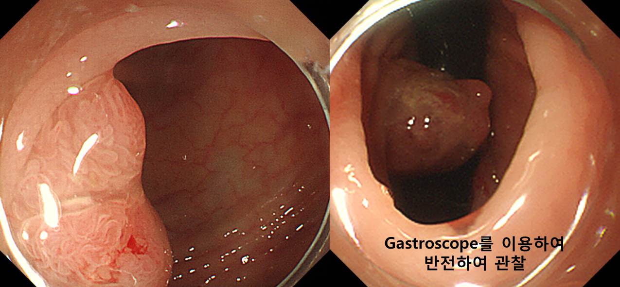
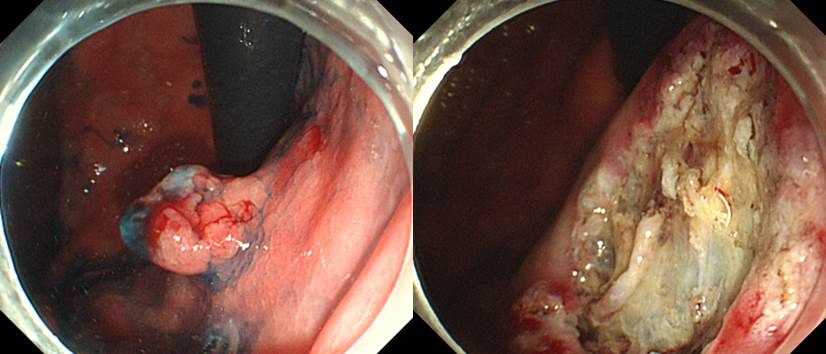
![]() 5. S-colon cancer
5. S-colon cancer
S colon의 병변인데 뒤쪽의 모양이 궁금했습니다. 일반 colonoscope로 반전이 어려워 위내시경을 이용하여 반전하여 관찰할 수 있었습니다. 무리하여 내시경 치료를 하지 않고 바로 수술을 결정하는데 도움이 되었습니다.
Adenocarcinoma, well differentiated
1. Location: sigmoid colon
2. Gross type: fungating
3. Size: 1.7x1.2 cm
4. Depth of invasion: invades submucosa (sm3)(pT1)
5. Resection margin: free from carcinoma, safety margin: proximal, 4 cm ; distal, 4.5 cm ; radial, >10 mm
6. Regional lymph node metastasis : No metastasis in all 25 regional lymph nodes(pN0)
7. Lymphatic invasion: not identified
8. Venous invasion: not identified
9. Perineural invasion: not identified
10. Tumor budding : negative
11. Micropapillary component: no
12. Tumor border: infiltrative
13. Pathologic staging: pT1 N0
![]() [References]
[References]
1) SMC Endoscopy Unit 삼성서울병원 내시경실
2) SMC Monday GI conference 삼성서울병원 일원내시경교실 월요점심소화기집담회
3) SMC Thursday endoscopy conference 삼성서울병원 일원내시경교실 목요점심내시경집담회
© 일원내시경교실 바른내시경연구소 이준행. EndoTODAY Endoscopy Learning Center. Lee Jun Haeng.