EndoTODAY 내시경 교실
Beginner | ESA | Schedule | OPD
Seminars | Atlas | Recent | Links
![]() [Thursday Endoscopy Conference 20170525]
[Thursday Endoscopy Conference 20170525]
![]() 1. Duodenal NET
1. Duodenal NET
개업가에서 십이지장 용종절제술 후 최종 결과가 neuroendocrine tumor로 나왔던 환자입니다. 추적관찰 중입니다.
Duodenum, polypectomy:
WELL DIFFERENTIATED NEUROENDOCRINE TUMOR (G1)
1) tumor size: 0.3x0.2 cm
2) extent: submucosa
3) lymphovascular invasion: not identified
4) perineural invasion: not identified
5) mitosis: 0/10 HPFs
6) resection margin; seems to be negative
. Synaptophysin: Positive in tumor cells
. Ki-67: Positive in 2% of tumor cells
* 참고: EndoTODAY 십이지장 NET
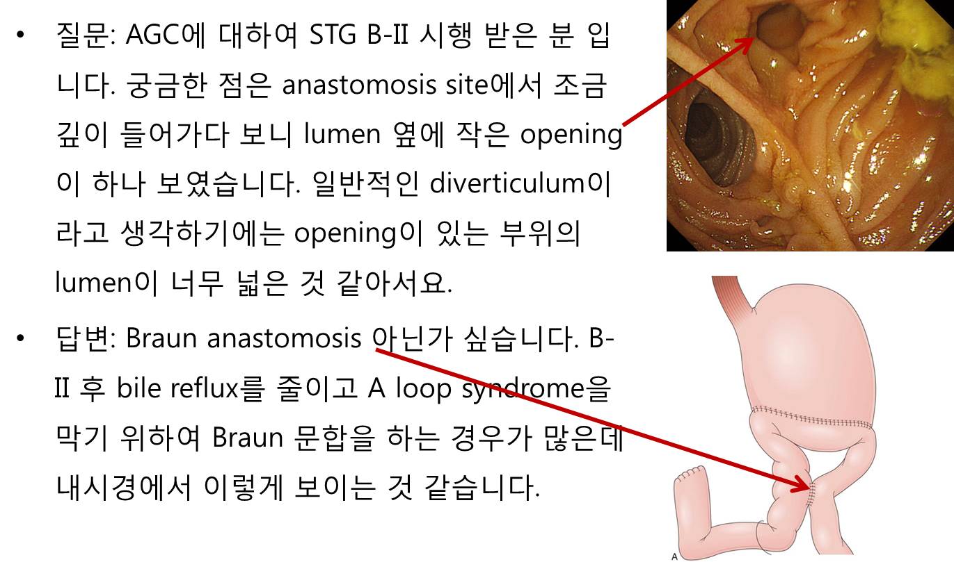
![]() 2. Braun anastomosis
2. Braun anastomosis
![]() 3. Borrmann type IV
3. Borrmann type IV
두 번 조직검사에서 암이 나오지 않았으나 육안소견 등 의거 수술을 시행하였습니다.
Stomach, total gastrectomy
Advanced gastric carcinoma
1. Location : [1] upper third, [2] middle third, Center at high body, anterior wall
2. Gross type : Borrmann type 4
3. Histologic type : undifferentiated carcinoma
4. Histologic type by Lauren : diffuse
5. Size : 13.5cm x encircled
6. Depth of invasion : invades serosa (pT4a)
7. Resection margin: free from carcinoma, safety margin: proximal 2 cm, distal 8 cm
8. Lymph node metastasis : metastasis to 2 out of 40 regional lymph nodes (pN1)
9. Lymphatic invasion : present
10. Venous invasion : not identified
11. Perineural invasion : present
12. Peritoneal cytology : negative
13. AJCC stage by 7th edition: T4a N1
* 참고: EndoTODAY 보만 4형 진행성 위암
PPT PDF 8.3 M
![]() 4. EGC after Ivor Lewis operation for esophageal cancer
4. EGC after Ivor Lewis operation for esophageal cancer
식도암이 발견되었습니다. 크기 및 깊이에 비하여 점막병소가 미약하였던 경우입니다. 이런 식도암은 놓치기 쉬운데, 검사자께서 참 잘 발견하신 경우입니다.

Esophagus and upper stomach, Ivor Lewis operation:
Invasive squamous cell carcinoma, moderately differentiated, distal esophagus:
1) tumor size: 4.5x3 cm
2) extension to perimuscular adventitia (pT3)
3) endolymphatic tumor emboli: not identified
4) perineural invasion: not identified
5) negative resection margins (proximal, 6.5 cm ; distal, 4 cm)
6) metastasis to 5 out of 52 regional lymph nodes (pN2) (5/52: "LC omentum", 0/0; "RRLN", 0/1; "LRLN", 0/6; "LD", 0/1; "5", 0/6; "7", 0/6; "8u", 0/2; "R9", 0/2; "L9", 0/1; "R10", 0/2; "L10", 0/4; "G1", 0/7; "G2", 0/2; "G3", 5/12)
식도암 치료는 잘 되었는데 6년 후 위암이 발견되었습니다. 매우 작았기 때문에 ESD로 치료하였습니다.
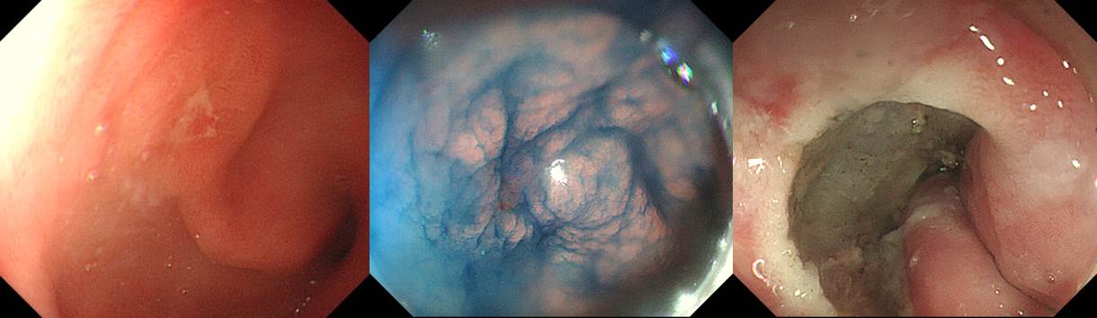
ESD. EGC
1. Location : antrum, anterior wall
2. Gross type : EGC type IIc
3. Histologic type : tubular adenocarcinoma, well differentiated
4. Histologic type by Lauren : intestinal
5. Size : (1) longest diameter, 6mm (2) vertical diameter, 6mm
6. Depth of invasion : invades mucosa (pT1a)
7. Resection margin: free from carcinoma (N), safety margin: proximal 10mm, distal 10mm, anterior 6mm, posterior 12mm
8. Lymphatic invasion : not identified
9. Venous invasion : not identified
10. Perineural invasion : not identified
11. Microscopic ulcer : absent
12. Histologic heterogeneity : absent
* 참고: EndoTODAY 식도암 후 위암
PDF, 0.2 M
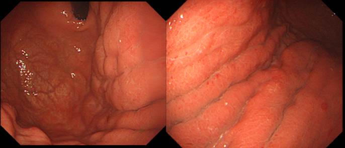
![]() 5. Rectal SMT
5. Rectal SMT
조직검사에서 nonspecific proctitis였고 EUS 후 내시경 절제술 하였고 GIST로 나왔습니다.
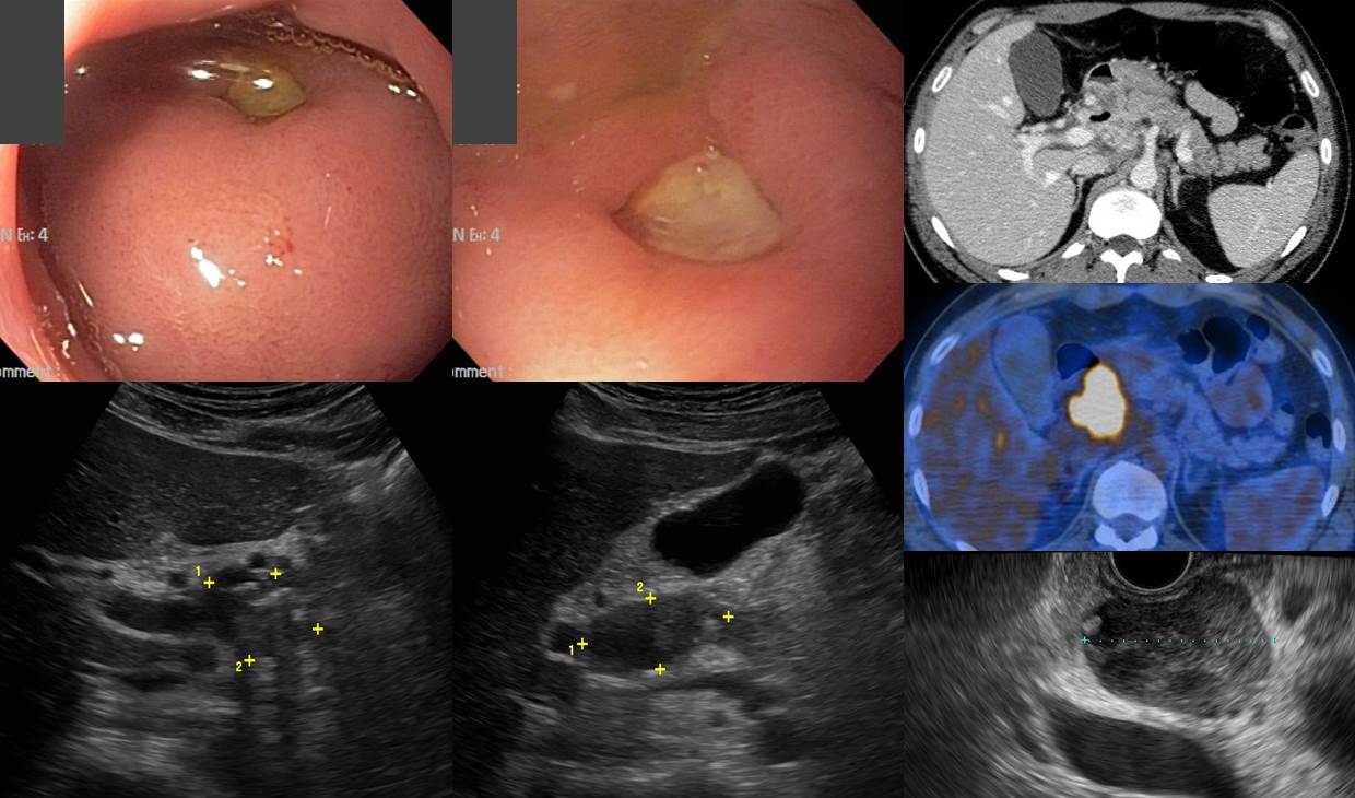
![]() 6. 복부 결핵성 림프절염의 십이지장 침윤
6. 복부 결핵성 림프절염의 십이지장 침윤
2개월 정도의 비특이적 상부위장관 불편감이 있던 40대 여성입니다. 내시경 검사에서 십이지장 구부의 둥근 융기부가 있고 그 중앙의 궤양이 발견되었고 같은 날 시행한 복부 초음파에서 췌장 주변의 mass가 의심되어 의뢰되었습니다. CT, PET, EUS-guided FNA 등을 시행하였습니다. CT에서는 multiple lymph adenopathy가 있고 십이지장 구부에 인접한 림프절 내부에 air가 보였습니다.
Abdominal TB lymphadenitis with fistula into the duodenal bulb로 항결핵제를 투여하였습니다. PPI가 필요한지 명확하지 않았습니다. 감염내과에서 PPI를 받아 드시고 계셨는데 중단을 권하지는 않았습니다.
* 참고: EndoTODAY 위장관 결핵
![]() [References]
[References]
1) SMC Endoscopy Unit 삼성서울병원 내시경실
2) SMC Monday GI conference 삼성서울병원 일원내시경교실 월요점심소화기집담회
3) SMC Thursday endoscopy conference 삼성서울병원 일원내시경교실 목요점심내시경집담회
© 일원내시경교실 바른내시경연구소 이준행. EndoTODAY Endoscopy Learning Center. Lee Jun Haeng.