EndoTODAY 내시경 교실
Beginner | ESA | Schedule | OPD
Seminars | Atlas | Recent | Links
![]() [Gastric cancer 432 - flat discolorated lesion]
[Gastric cancer 432 - flat discolorated lesion]
001 | 101 | 201 | 301 | 401 | 501 | 601 | 701 | 801 | 901 | 1000
저는 단순한 것을 좋아합니다. 제 모토는 이것입니다. Use simple tools very well! 하지만 단 하나의 도구로 모든 상황을 해결하기는 어렵습니다. 망치가 필요할 때도 있고 도끼가 필요할 때도 있는 법입니다.
위의 함몰형 병소가 관찰되면 양성 궤양과 악성 궤양의 감별이 중요한 포인트입니다. 궤양의 양성/악성 감별을 위해서는 fold, edge, margin, base 소견을 잘 살펴야 합니다.
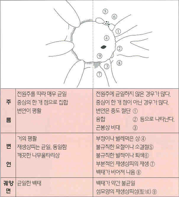
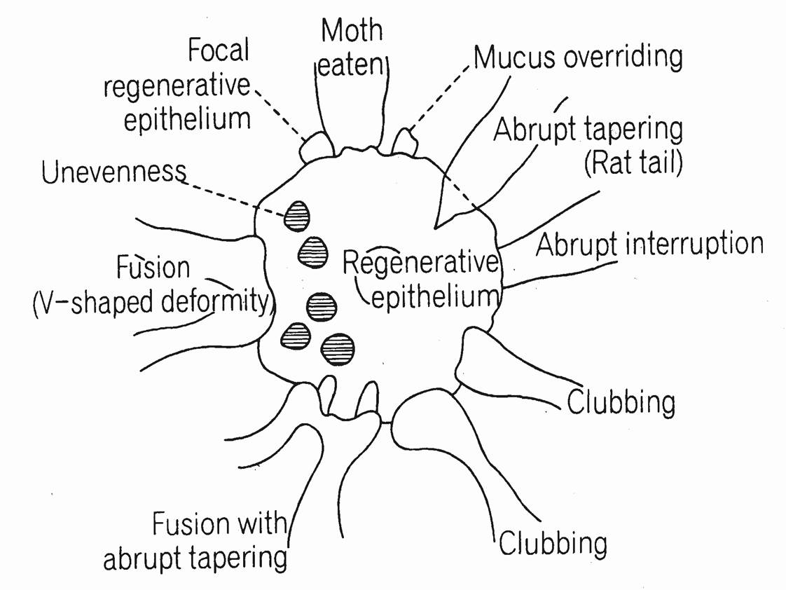
악성궤양의 주름은 궤양 바닥에서 떨어진 곳에서 끝나거나 abrupt cutting (=interruption), rat-tailing (=rapid tapering), clubbing, fusion, dam-formation 등을 보입니다. 악성궤양의 또 다른 소견은 edge와 margin에서 관찰할 수 있는데, 궤양변연의 뚜렷한 결절 모습, 주변 점막의 불규칙한 변색, 부분적인 재생상피, 백태의 overriding, 위 소구의 패턴의 소실 등입니다. 악성 궤양의 바닥은 불규칙한 경향이 있으나 평탄해 보이는 경우도 있습니다. 궤양내의 tumor island도 중요한 소견입니다. 반대로 양성 궤양의 바닥은 상대적으로 smooth하며 궤양의 경계가 명확하고 위에서 언급된 악성을 시사하는 소견이 없습니다. 양성 궤양의 주름은 궤양 바닥에서 끝나는 경향이 있고 궤양이 큰 경우에는 변연에 흡수되기도 합니다.
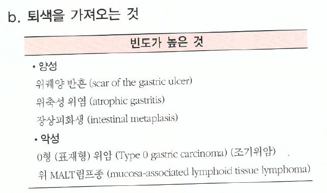
Flat (or slightly depressed) discolorated lesion에서는 다릅니다. 궤양 감별에 사용되는 fold, edge, margin, base 소견을 flat (or slightly depressed) discolorated lesion에 적용하기는 어렵습니다. 뚜렷하지도 않은 fold, edge, margin, base 소견을 과장하여 기술한들 도움이 되지 않습니다. 그냥 위의 'Flat (or slightly depressed) discolorated lesion 감별진단 table'을 떠올리면서 어느 것이 가장 비슷한지 전후좌우를 살펴보는 것이 낫습니다.
'상부소화관 내시경 소견의 판독 및 감별진단 (박인서 번역)' 140 쪽에는 작은 퇴색형 병소의 감별진단 table이 제시되어 있습니다. Fold, edge, margin, base를 생각하기보다는 이 table을 떠올리는 편이 접근하기 쉽습니다.

BGU scar
국소적 위축성 위염
화생성 위염
EGC IIb (poorly differentiated adenocarcinoma)
EGC IIb (SM1 invasion, signet ring cell carcinoma)
Stomach, endoscopic submucosal dissection:
Early gastric carcinoma
1. Location : proximal antrum, greater curvature
2. Gross type : EGC type IIb
3. Histologic type : tubular adenocarcinoma, moderately differentiated
4. Histologic type by Lauren : intestinal
5. Size : 2.2x1.5x0.05 cm
6. Depth of invasion : invades mucosa (lamina propria) (pT1a)
7. Resection margin: free from carcinoma, safety margin: distal 1 cm, proximal 0.6 cm, anterior 1 cm, posterior 0.6 cm
8. Lymphatic invasion : not identified
9. Venous invasion : not identified
10. Perineural invasion : not identified
11. Microscopic ulcer : absent
12. Histologic heterogeneity: absent
MALToma
MALToma
상부소화관 내시경 소견의 판독 및 감별진단 (박인서 번역) 140-141 쪽을 scan 하였습니다. 참고하시기 바랍니다.
PDF 0.9 M
옛날 책이 도움될 때가 있습니다. 특히 옛날 일본 책 번역본은 크게 도움이 됩니다. 일본 내시경 책 중 가장 좋은 책들만 번역되었다고 생각해도 좋습니다.
© 일원내시경교실 바른내시경연구소 이준행. EndoTODAY Endoscopy Learning Center. Lee Jun Haeng.