EndoTODAY 내시경 교실
Beginner | ESA | Schedule | OPD
Seminars | Atlas | Recent | Links
 [Gastric cancer 508]
001 | 101 | 201 | 301 | 401 | 501 | 601 | 701 | 801 | 901 | 1000
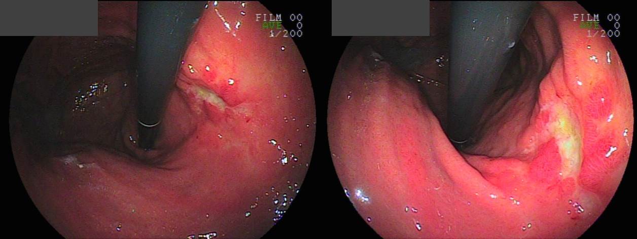
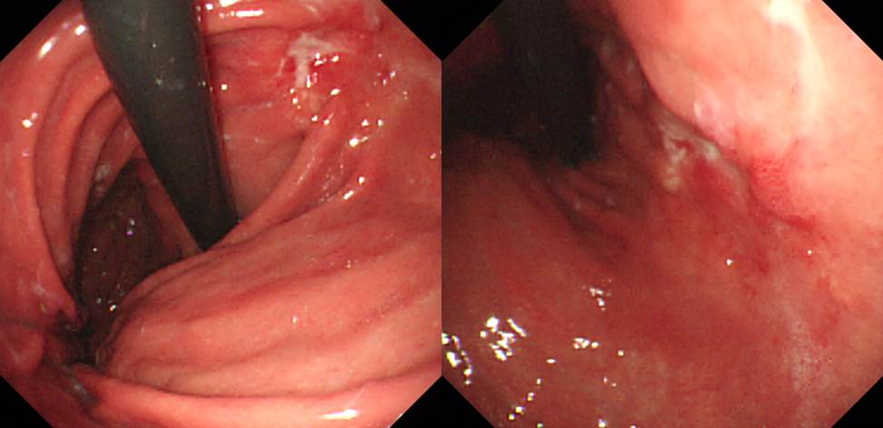
내시경에서 암이 발견되어 의뢰된 경우 위암의 모양이 상당히 달라져서 놀라는 예가 있습니다. 보통 궤양형 암에서 PPI나 H2RA를 복용하신 환자들입니다.
아래는 의뢰 후 재검 시 사진입니다. 많이 달라졌습니다.
수술 후 병리는 아래와 같았습니다.
- Histologic type : tubular adenocarcinoma, poorly (poorly cohesive) differentiated
- Size : 4.8x4.1 cm
- Depth of invasion : penetrates subserosal connective tissue (pT3)
- Resection margin: free from carcinoma
- Lymph node metastasis : metastasis to 4 out of 45 regional lymph nodes (pN2)
- Lymphatic invasion : present (+++)
- AJCC stage by 7th edition: pT3 N2
© 일원내시경교실 바른내시경연구소 이준행. EndoTODAY Endoscopy Learning Center. Lee Jun Haeng.