EndoTODAY 내시경 교실
Beginner | ESA | Schedule | OPD
Seminars | Atlas | Recent | Links
 [Gastric cancer 537 - 병소의 범위 파악이 매우 어려웠던 보만 4형 진행성 위암]
001 | 101 | 201 | 301 | 401 | 501 | 601 | 701 | 801 | 901 | 1000
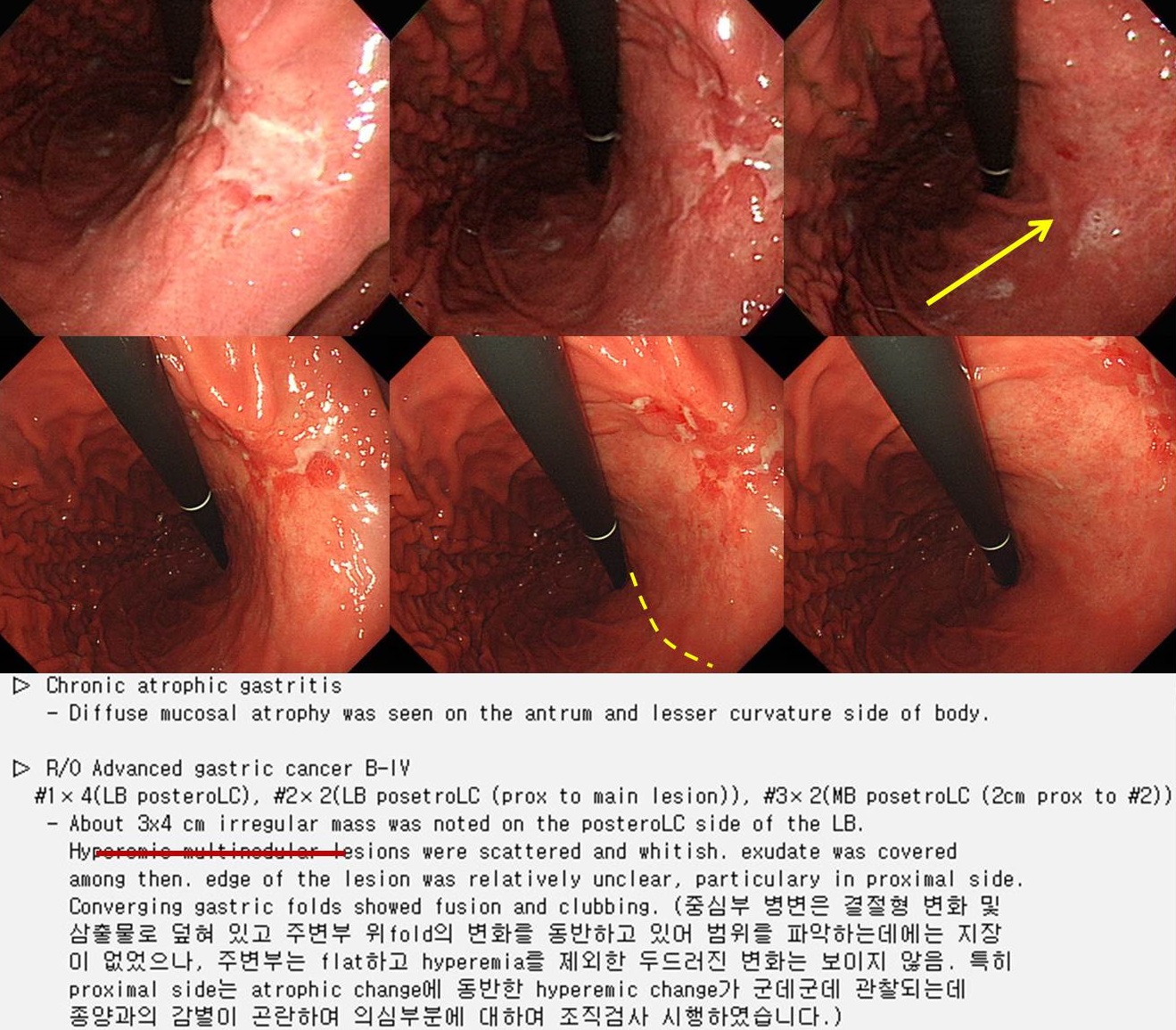
Signet ring cell carcinoma의 조직진단으로 의뢰된 환자입니다. 위각 직상방에 경계가 불규칙한 함몰형 병소가 있었으나 그 근위부로 널고 불규칙한, 군데군데 발적을 동반한 atrophic area가 관찰되어 병소의 proximal margin 판단이 어려웠습니다. 재검을 하였습니다.
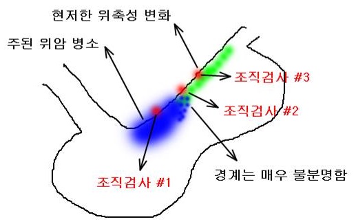
한눈에 잘 보이는 함몰부 상방의 암침윤이 의심되었으므로 일견 의심되는 병소의 proximal margin 보다 1 cm 근위부 (biopsy #2)와 그보다 2cm 근위부(biopsy #3)에서 조직검사를 했는데 모두 암으로 확인되었습니다.
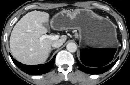
CT에서 위벽은 위체부 소만쪽으로 아주 조금 두꺼워진 정도였습니다.
Total gastrectomy를 시행할 수 밖에 없었는데 병리결과는 예상보다 훨씬 심했습니다.
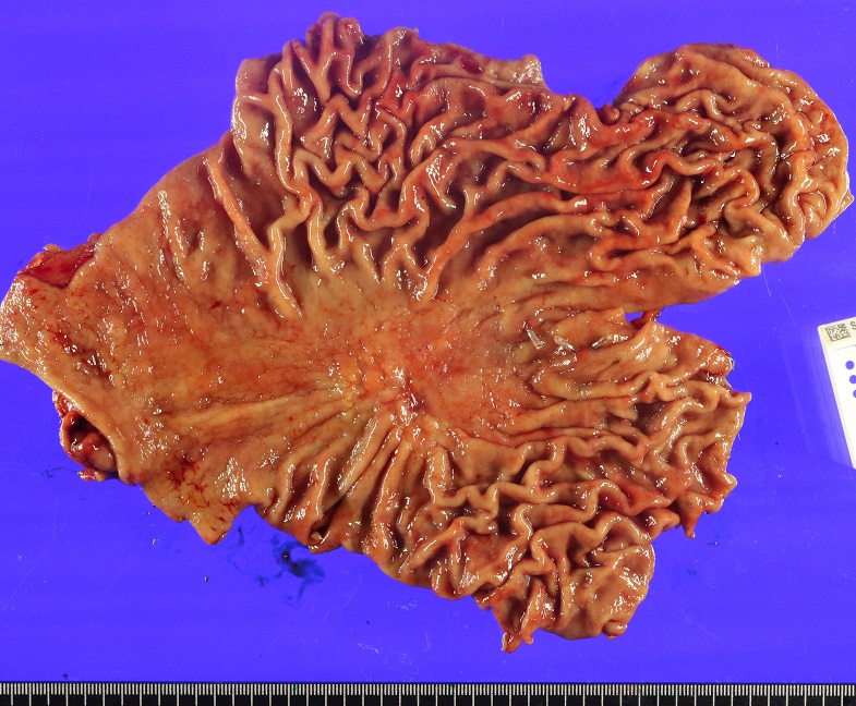
Stomach, total gastrectomy:
. Advanced gastric carcinoma
1. Location : upper third, Center at cardia (Siewert III) and lesser curvature
2. Gross type : Borrmann type 4
3. Histologic type : tubular adenocarcinoma, poorly (poorly cohesive) differentiated
4. Histologic type by Lauren : diffuse
5. Size : 5x3.7 cm
6. Depth of invasion : penetrates subserosal connective tissue (pT3)
7. Resection margin: free from carcinoma, safety margin: proximal 1.5 cm, distal 7 cm
8. Lymph node metastasis : metastasis to 3 out of 38 regional lymph nodes (pN2) (perinodal extension: present) (3/38: "2", 0/4; "3", 2/4; "4", 0/4; "5", 0/0; "6", 0/5; "7", 0/2; "9", 1/10; "8a", 0/2; "11p", 0/1; "12a", 0/3; "4sb", 0/0; "1", 0/2; "perigastric LN", 0/1)
9. Lymphatic invasion : not identified
10. Venous invasion : not identified
11. Perineural invasion : present
12. AJCC stage by 7th edition: pT3 N2
미분화 조직형 위암의 경계판정은 매우 어렵습니다만, 이처럼 함몰부 주위 넓게 발적 퇴색된 병소를 동반한 경우는 어디까지 암인지 어디까지 위축성 변화인지 짐작하기란 매우 어려운 일이라고 생각되었습니다. 첫 사진을 다시 곰곰히 살펴본 바 fold가 끊긴 부분(화살표)이 암과 정상의 경계였습니다. 검사를 잘 해주신 fellow 선생님께 감사드립니다.
© 일원내시경교실 바른내시경연구소 이준행. EndoTODAY Endoscopy Learning Center. Lee Jun Haeng. (2017-10-23)