EndoTODAY 내시경 교실
Beginner | ESA | Schedule | OPD
Seminars | Atlas | Recent | Links
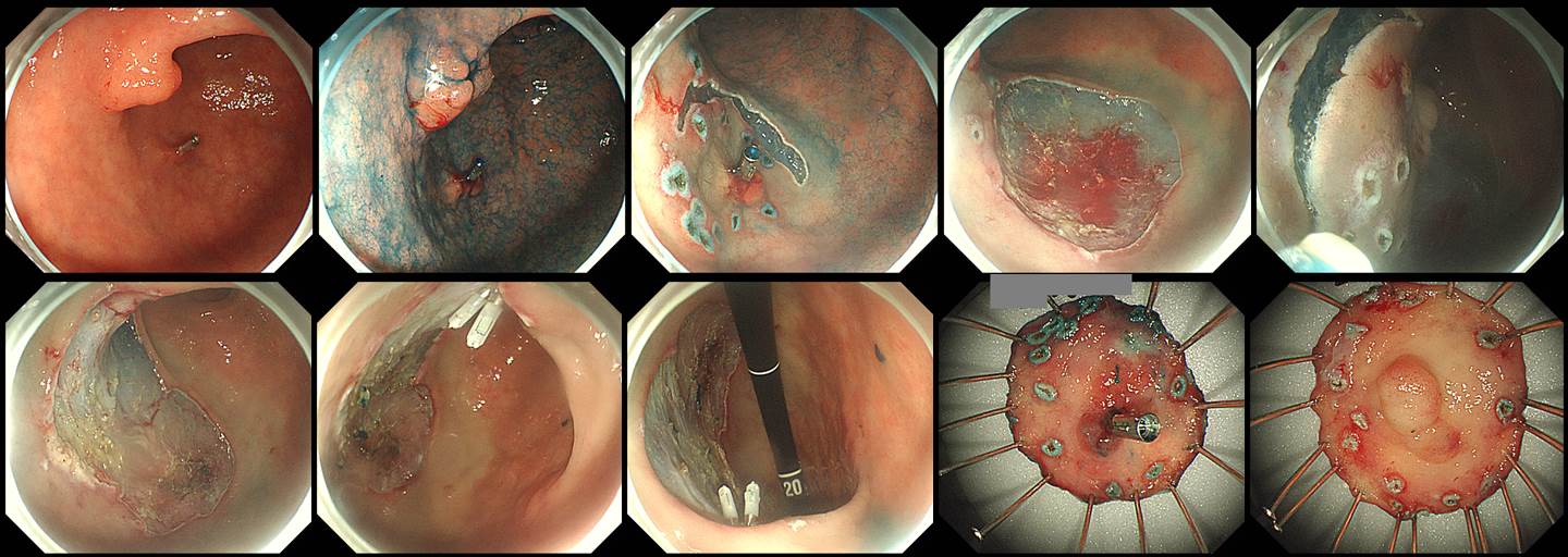
![]() [Gastric cancer 779. Two EGCs]
[Gastric cancer 779. Two EGCs]
001 | 101 | 201 | 301 | 401 | 501 | 601 | 701 | 801 | 901 | 1000
Two neoplastic lesions were found during screening gastroscopy.
Pentax EPK-i5000 system + EG-2990i endoscope
When two EGCs are located very closely, would you do ESD in one piece (plan A)or in two pieces (plan B)?
Plan A and B
I usually choose the plan B (two pieces strategy). If I choose the plan A, it is considered as one ESD procedure. If I choose the plan B, it is considered as two ESD procedures. Reimbursement issue is something that I cannot ignore.
One concern regarding the plan B is that the resection margin of both specimen is positive at the same time. It means it was actually one cancer rather than two closeby cancers.
Anyway, the final pathology was as follows;
Antero-GC of proximal antrum, ESD: Early gastric carcinoma
1. Location : proximal antrum, antero-lesser curvature
2. Gross type : EGC type IIa
3. Histologic type : tubular adenocarcinoma, well differentiated
4. Histologic type by Lauren : intestinal
5. Size of carcinoma : (1) longest diameter, 8 mm (2) vertical diameter, 7 mm
6. Depth of invasion : invades mucosa (lamina propria) (pT1a)
7. Resection margin : free from carcinoma(N), safety margin : distal 9 mm, proximal 10 mm, anterior 10 mm, posterior 6 mm, deep 400
8. Lymphatic invasion : not identified(N)
9. Venous invasion : not identified(N)
10. Perineural invasion : not identified(N)
11. Microscopic ulcer : absent
12. Histologic heterogeneity: absentAnterior wall of angle, ESD: Early gastric carcinoma
1. Location : angle, anterior wall
2. Gross type : EGC type IIa
3. Histologic type : tubular adenocarcinoma, well differentiated
4. Histologic type by Lauren : intestinal
5. Size of carcinoma : (1) longest diameter, 10 mm (2) vertical diameter, 10 mm
6. Depth of invasion : invades mucosa (lamina propria) (pT1a)
7. Resection margin : free from carcinoma(N), safety margin : distal 7 mm, proximal 7 mm, anterior 20 mm, posterior 8 mm, deep 700
8. Lymphatic invasion : not identified(N)
9. Venous invasion : not identified(N)
10. Perineural invasion : not identified(N)
11. Microscopic ulcer : absent
12. Histologic heterogeneity: absent
13. Associated finding: Gastritis cystica superficialis
© 일원내시경교실 바른내시경연구소 이준행. EndoTODAY Endoscopy Learning Center. Lee Jun Haeng. (2019-8-10)