EndoTODAY 내시경 교실
Beginner | ESA | Schedule | OPD
Seminars | Atlas | Recent | Links
![]() [Gastric cancer 840. Referred due to adenoma, but an EGC was found and ESDed.]
[Gastric cancer 840. Referred due to adenoma, but an EGC was found and ESDed.]
001 | 101 | 201 | 301 | 401 | 501 | 601 | 701 | 801 | 901 | 1000
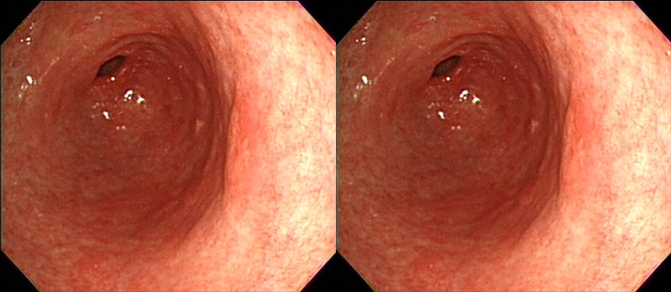
70 years old lady was referred for the endoscopic treatment of a small pale elevated adenoma of the posterior antral wall.
In the ESD procedure, I could not find the index lesion (a small pale elevated adenoma of the posterior antral wall). Instead, a small depressed EGC-like lesion was found in the GC side of the mid-antrum. ESD was done for the depressed lesion and APC ablation was done for suspeced white lesions in the posterior antral wall.

ESD: Early gastric carcinoma
1. Location : antrum, greater curvature and posterior wall
2. Gross type : EGC type IIc
3. Histologic type : tubular adenocarcinoma, well differentiated
4. Histologic type by Lauren : intestinal
5. Size of carcinoma : (1) longest diameter, 10 mm (2) vertical diameter, 9 mm
6. Depth of invasion : invades mucosa (muscularis mucosa) (pT1a)
7. Resection margin : free from carcinoma(N), safety margin : distal 8 mm, proximal 11 mm, anterior 10 mm, posterior 16 mm, deep 500 ㎛
8. Lymphatic invasion : not identified(N)
9. Venous invasion : not identified(N)
10. Perineural invasion : not identified(N)
11. Microscopic ulcer : absent
12. Histologic heterogeneity: absent
In summary, I think it is a synchronous cancer lesion in a patient referred for the adenoma. Careful examination of the whole stomach mucosa is important before ESD.
이번 case의 제목을 "840. Referred due to adenoma, but an EGC was found and ESDed."로 잡아보았습니다. 맨날 ESD를 하다보니 이제는 ESD가 동사처럼 느껴집니다.
© 일원내시경교실 바른내시경연구소 이준행. EndoTODAY Endoscopy Learning Center. Lee Jun Haeng. (2020-3-16)