 EndoTODAY 내시경 교실
Beginner | ESA | Schedule | OPD
Seminars | Atlas | Recent | Links
![]() [Killian-Jamieson diverticulum]
[Killian-Jamieson diverticulum]
2020-4-25. 순천만내시경세미나 동영상 강의
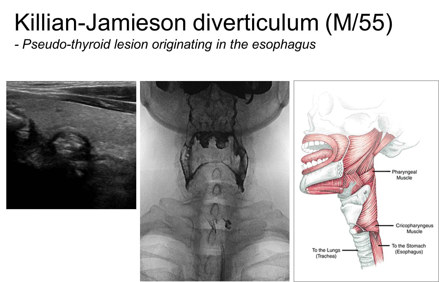
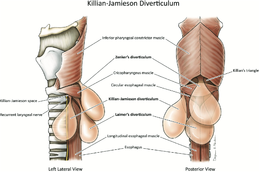
건강검진 갑상선 초음파검사에서 식도 병변이 갑상선 nodule로 오인되어 내분비내과로 의뢰되는 경우가 발생하고 있습니다. 이 환자는 Killian-Jamieson diverticulum로 진단되었습니다. 감별해야 할 것은 Zenker's diverticulum입니다. 가장 현저한 차이점은 Killian-Jamieson diverticulum는 cricopharyngeus의 아래 level, anterolateral aspect에 발생하고 Zenker’s diverticulum은 cricopharyngeus의 윗 level, posterior aspect에서 발생한다는 점입니다.
Zenker's diverticulum originates on the posterior wall of the pharyngoesophageal segment in a midline area of weakness just above the cricopharyngeus (i.e., Killian's dehiscence), whereas Killian-Jamieson diverticula originate on the anterolateral wall of the proximal cervical esophagus in a gap just below the cricopharyngeus and lateral to the longitudinal tendon of the esophagus (i.e., the Killian-Jamieson space).
The cricopharyngeus muscle is located at the junction of the pharynx (throat) and esophagus, and is the major muscular component of what is called the upper esophageal sphincter (UES). At rest, the UES closes the passageway between the pharynx and esophagus.
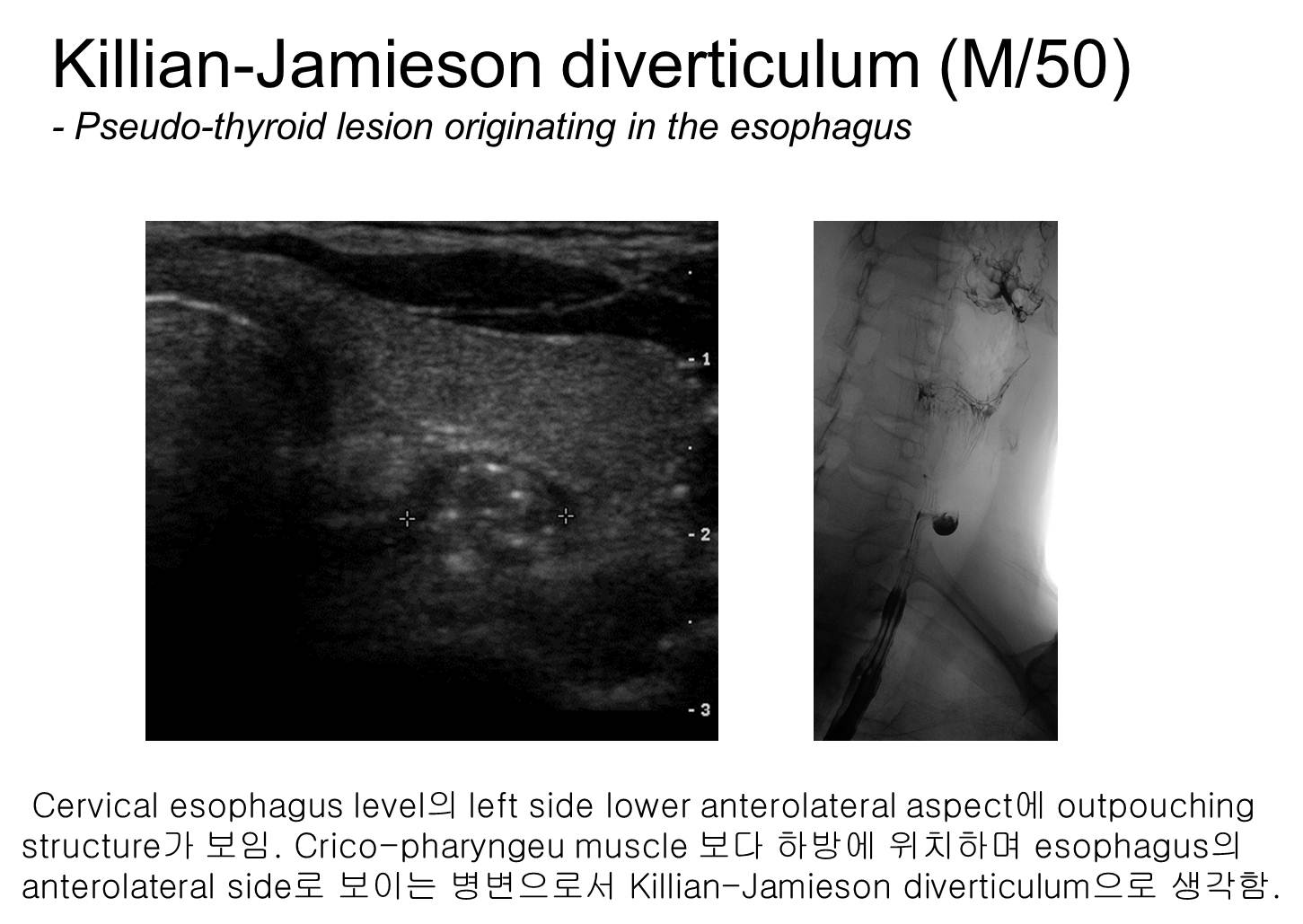

간혹 KJ diverticulum은 갑상선 초음파에서 갑상선 암으로 오인될 수 있습니다. 갑상선 암 의심하에 aspiration을 하게 되면 tumor가 나오지 않고 food material이 나와서 깜짝 놀라기도 합니다.
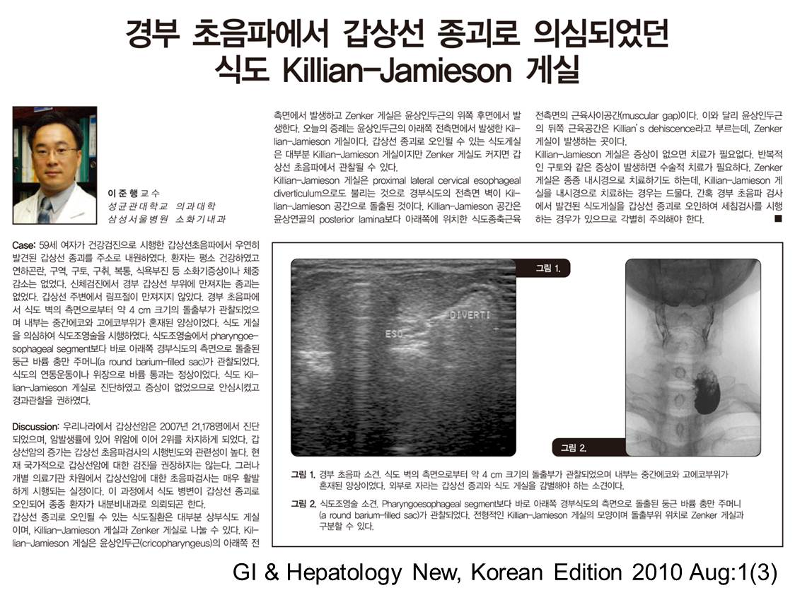
증상이 없어서 경과관찰만 권하였습니다.
* EndoTODAY 애독자 증례 편지 49 - KJ diverticulum
![]() [References]
[References]
1) 식도 게실: Zenker diverticulum, Killian-Jamieson 게실, 중부식도 게실, 하부식도 게실
2) 위 게실
3) 십이지장 게실
4) Meckel 게실
5) 대장 게실과 게실염
© 일원내시경교실 바른내시경연구소 이준행. EndoTODAY Endoscopy Learning Center. Lee Jun Haeng.