EndoTODAY 내시경 교실
Beginner | ESA | Schedule | OPD
Seminars | Atlas | Recent | Links
![]() [중부식도게실. Mid-esophageal diverticulum] - 終
[중부식도게실. Mid-esophageal diverticulum] - 終
2020-4-25. 순천만내시경세미나 동영상 강의
![]() 1. 내시경 소견
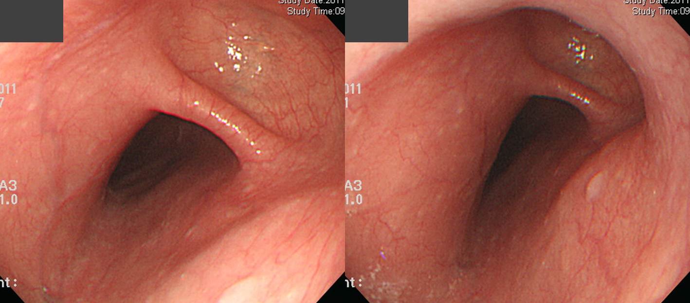
1. 내시경 소견
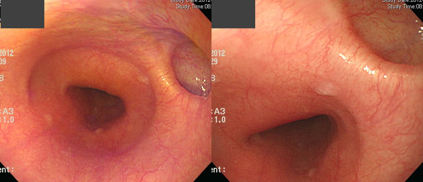
Pentax Imagina로 촬영한 중부식도 게실
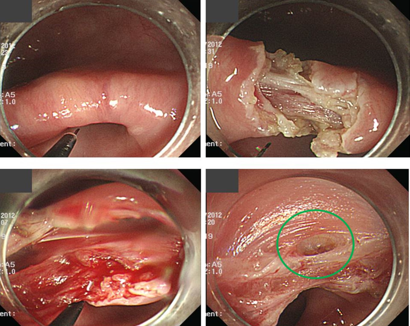
![]() 2. 중부식도게실 내시경치료
2. 중부식도게실 내시경치료
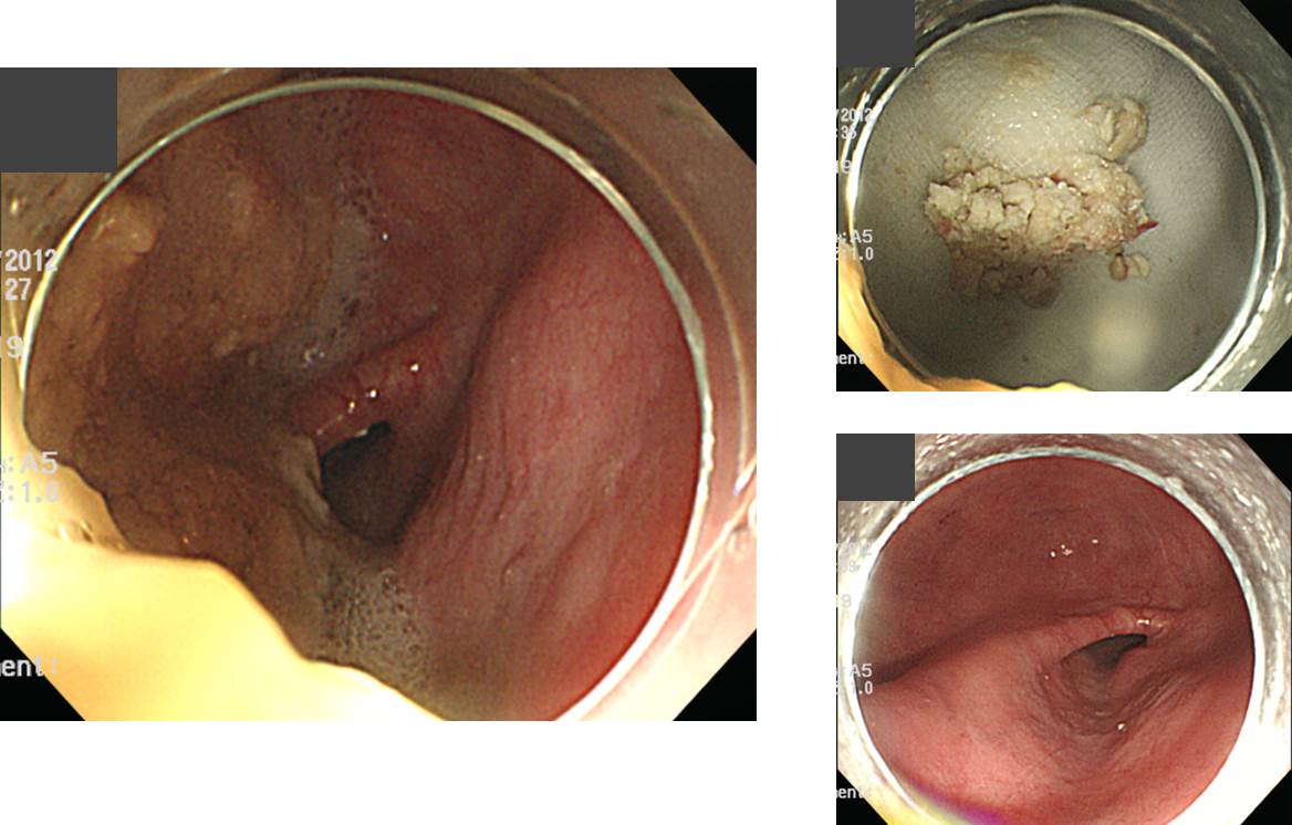
중부식도게실이 크면 증상이 발생할 수 있습니다. 초고령 환자였습니다. 수술 이외의 다른 방법이 없는데, 수술은 너무 위험하니 음식을 조금씩 드시고 물을 많이드시라는 이야기만 들었다고 합니다. 2차 의견을 위하여 방문하였습니다.
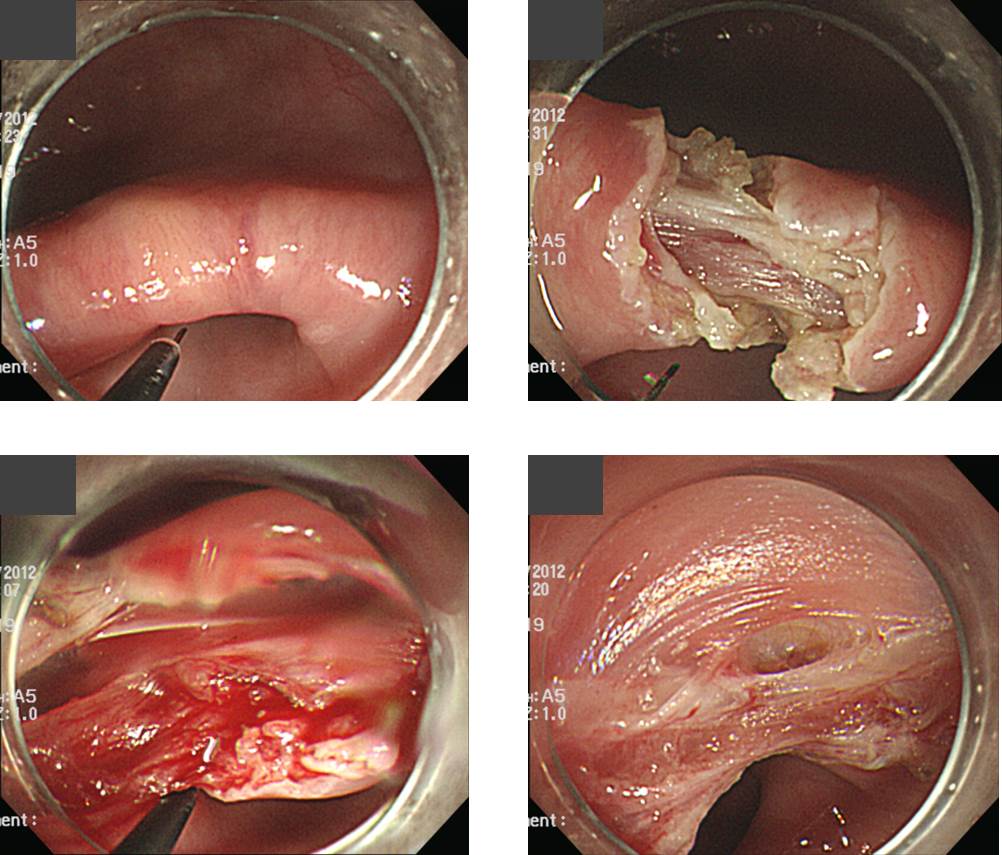
Endoscopic septal dissection을 시도하기로 하고 Zenker diverticulum 내시경치료 하는 것과 비슷하게 knife를 이용하여 septum을 절개하였습니다. 증상은 즉시 좋아졌습니다. 천공이 걱정이지만 시술은 그리 어렵지 않았습니다.
![]() 3. Midesophageal traction diverticulum with anthracotic pigmentation
3. Midesophageal traction diverticulum with anthracotic pigmentation
[2018-6-26. 애독자 질문]
이준행교수님께
안녕하세요? 늦었지만 작년에 위내시경 hands on 교육을 받을 수 있게 해주셔서 다시 한번 감사드립니다. 저도 모르게 익숙해진 내시경 습관들을 객관적으로 봐주시고 바로잡아 주셔서 이전보다 훨씬 안정적으로 검사할 수 있게 된 것 같습니다. 물론 스킬보다는 병변을 알아보는 내공이 중요하겠지요? 앞으로도 EndoTODAY를 통해 더 열심히 공부하겠습니다.
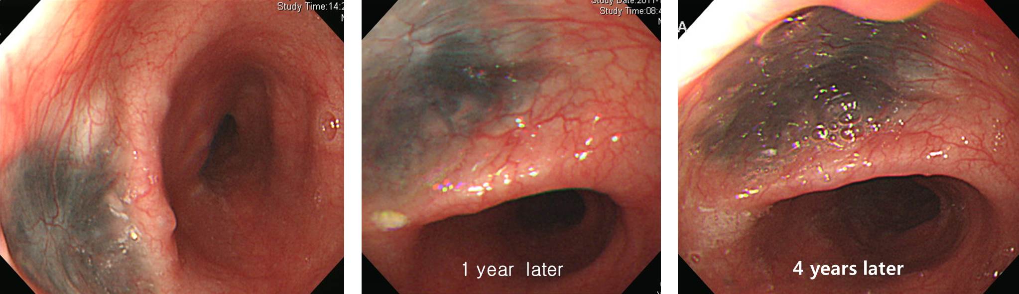
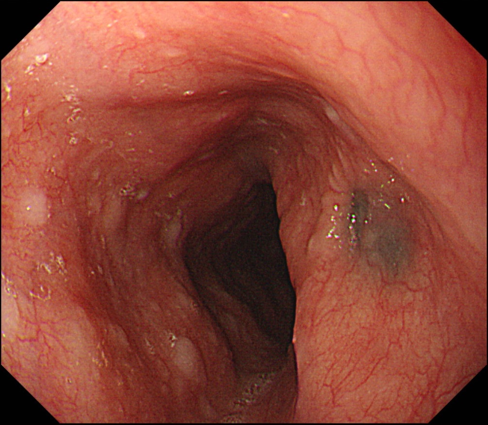
최근 비슷한 두 식도병변을 접하게 되어 문의드리고자 합니다. 중부식도 게실 기저면에 검푸른 색이 비춰 보이는 것으로 혹시 부식성 식도염의 scar tissue인가 싶어서 병력을 청취하였으나 특별한 점은 없었습니다.
증례 1
증례 2
교수님의 의견은 어떠신지요?
[2018-6-26. 이준행 답변]
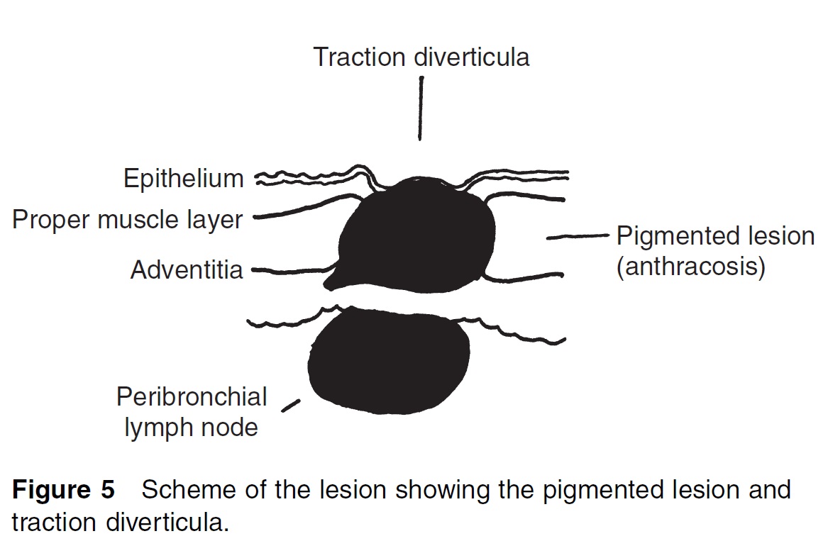
Traction diverticulum 바닥의 anthracotic pigmentation입니다. 우리나라에서 중부식도 traction diverticulum의 흔한 원인은 결핵성 mediastinal lymphadenopathy입니다. 결핵이 좋아지면서 식도 벽이 당겨져서 traction diverticulum이 만들어지는 것입니다.
결핵 환자의 기관지 내시경에서 anthracotic pigmentation이 자주 관찰되는 것은 잘 알고 계실 것입니다. 마찬가지로 결핵성 림프절염에서도 anthracotic pigmentation이 발생합니다. 결핵성 림프절염이 식도벽으로 파급하고 또 아물면서 식도벽이 당겨져 traction diverticulum이 되는 것인데, 이 과정에서 게실 바닥에 anthracotic pigmentation이 남게 됩니다.
아래는 제가 경험한 증례입니다.
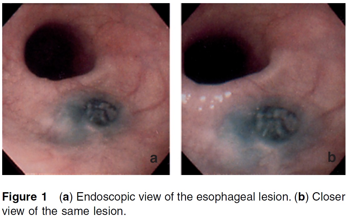
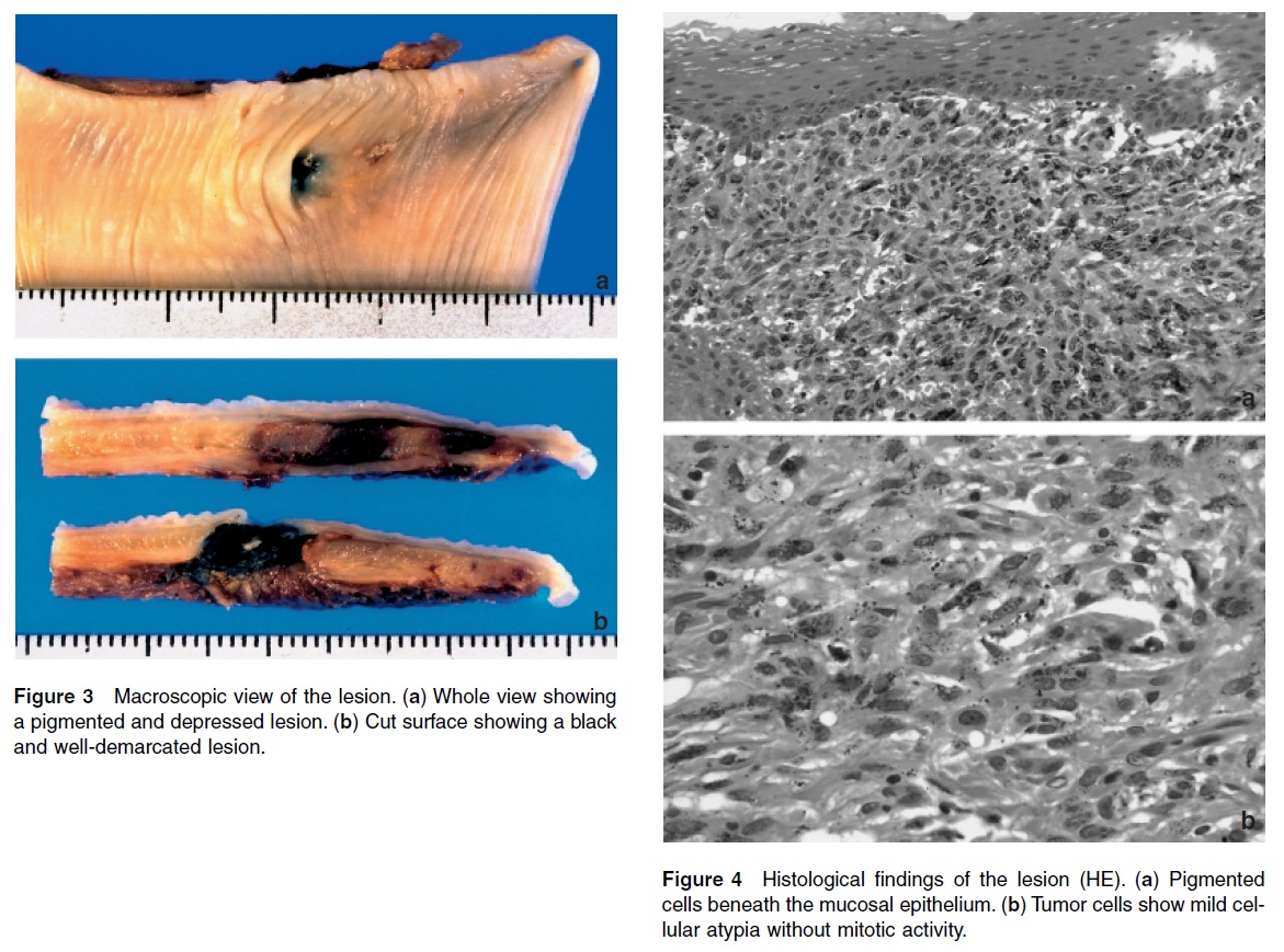
Pathology International 2002에 매우 인상적인 일본 증례 보고가 있어서 초록과 사진을 소개합니다.
"A case of anthracosis of the esophagus is reported. The patient was a previously healthy 69-year-old Japanese woman. A black and slightly elevated lesion was detected in her esophagus by upper gastroesophageal fiberoscopic examination. Endoscopically, the lesion looked like malignant melanoma. Thoracic esophagotomy was then performed. Histological examination revealed a pigmented lesion beneath the mucosal epithelial layer. The lesion consisted of an aggregation of histiocytes containing an abundance of tiny black pigments. A few mature lymphocytes and plasma cells were also evident in the periphery of the lesion. Histologically, these findings looked like lymph nodes in the pulmonary hilus; however, no lymph nodal structure was evident in the esophageal wall. Traction diverticula were also noted in the pigmented lesion. The patient has remained well without disease for 9 months since the surgery. Although anthracosis is a rare condition in the esophagus, the present case gave warning to pathologists and clinicians that it does indeed occur. Endoscopists and pathologists should differentiate anthracosis from malignant melanoma because the treatment and outcome are quite different for each."
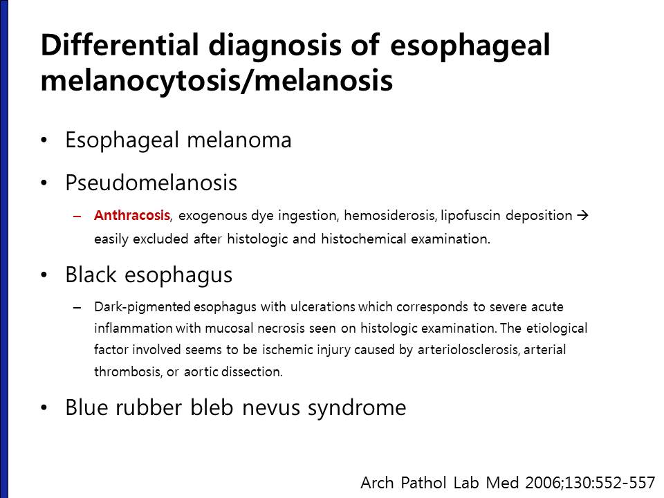
식도의 melanocytosis/melanosis와 감별해야 할 것 중 하나가 anthracosis입니다.
[2022-2-3. 애독자 질문]
연휴 동안 Esophageal diverticulum과 관련한 시술을 EndoToday에서 찾아보게 되었습니다. 이 중, 중부게실에서 septotomy (FESD)를 시행한 동영상과 사진을 매우 흥미롭게 보았습니다.
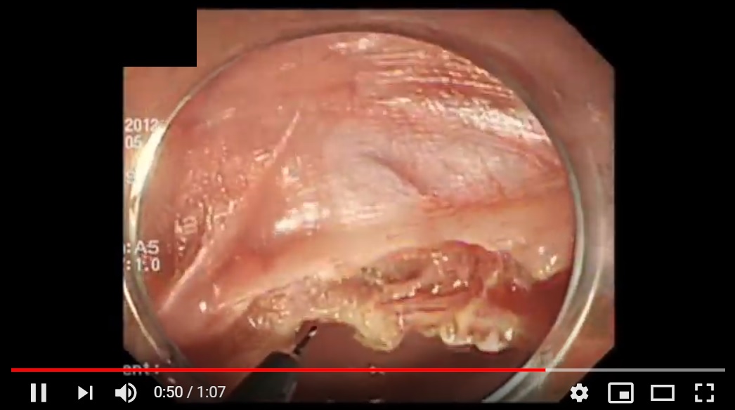
특히, 시술과 관련한 설명에서, septum을 거의 '빵구'가 날 정도로 깊게 쳐야 하고, 시술 시간은 15분 남짓으로 짧았으며, 천공의 위험성이 높다는 내용이 인상적이었습니다. 시술 동영상의 사진에서 초록색 원 부분이 좀 아슬아슬해 보였습니다만, 별도의 clipping을 하지 않고 종료한 것 같았습니다. 초록색 원 부분은 serosa가 노출된것은 아닌지 궁금하여 여쭙습니다.
아울러, '어느 정도'까지 septum을 쳐야 하는지 혹시 교수님만의 기준이나 landmark가 있으실지 여쭙습니다.
[2022-2-6. 이준행 답변]
답이 늦었습니다. 죄송합니다. 새해 복 많이 받으세요.
그렇습니다. 지적하신 바와 같이 시술하면서 초록생 동그라미 부위를 보고 "혹시 천공인가?" 걱정했는데 시술 후 흉부 사진에서 별 문제가 없고 환자도 특별한 불편감 없이 잘 회복하셨습니다. 제주도 환자였는데 증상이 좋아졌다고 기뻐하면서 저에게 천리향 한 박스를 보내주셨던 기억이 납니다. 김영란법 이전의 미담이지만...
아시는 바와 같이 시술과 관련된 천공은 대부분 금식과 항생제로 해결할 수 있습니다. 천공을 모르고 식사를 하여 종격동염이 발생한 경우가 문제이지 시술 후 적절히 살피고 충분히 금식하면 해결이 됩니다. 그래서 그리 걱정할 것은 없습니다. 증상이 재발되지 않도록 근육 fiber를 끝까지 끊어주는 것이 중요할 것 같습니다. 사실 초록색 동그라니 하단의 transverse한 근육층을 조금 더 cutting 할까말까 몇 번 망설였던 기억이 납니다. 항상 가능한 것은 아니지만 보이는 transverse한 근육층을 모두 cutting 하는 것이 일차적인 목표입니다.
저는 아직 시도한 적이 없지만 POEM 방법을 적용하면 보다 안전할 것 같기는 합니다. 앞으로는 비슷한 환자가 오시면 민양원 교수님이나 이혁 교수님께 의뢰하려고 합니다.
![]() [References]
[References]
1) 식도 게실: Zenker diverticulum, Killian-Jamieson 게실, 중부식도 게실, 하부식도 게실, 2022년 신철민 교수님 종설
2) 위 게실
3) 십이지장 게실
4) Meckel 게실
5) 대장 게실과 게실염
© 일원내시경교실 바른내시경연구소 이준행. EndoTODAY Endoscopy Learning Center.