 EndoTODAY 내시경 교실
Beginner | ESA | Schedule | OPD
Seminars | Atlas | Recent | Links
 [위암 108]
001 | 101 | 201 | 301 | 401 | 501 | 601 | 701 | 801 | 901 | 1000
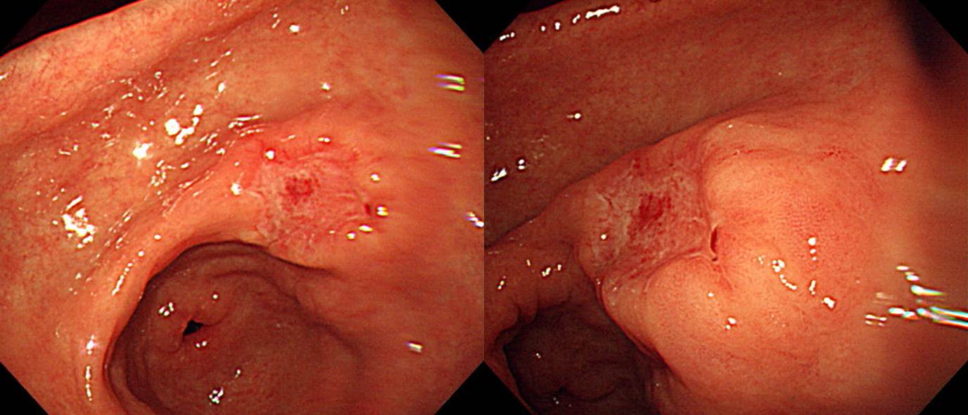
아래 두 사진 중 어느 것이 AGC이고 어느 것이 EGC일까요? 아래 두 사진 중 어느 것이 림프절 전이가 있고 어느 것이 림프절 전이가 없을까요?

아래로
아래로
아래로
아래로
첫번째 증례의 결과입니다.
Early gastric carcinoma
1. Location : lower third, Center at antrum and greater curvature
2. Gross type : EGC type IIc
3. Histologic type : tubular adenocarcinoma, poorly differentiated
4. Histologic type by Lauren : diffuse
5. Size : 3.2x1.6 cm
6. Depth of invasion : invades submucosa (sm3) (pT1b)
7. Resection margin: free from carcinoma, safety margin: proximal 7.3 cm, distal 5.3 cm
8. Lymph node metastasis : metastasis to 6 out of 47 regional lymph nodes (pN2), (perinodal extension: absent) (6/47: "1", 0/0; "4sb", 0/0; "5", 0/1; "6", 0/5; "8a", 0/2; "7", 0/4; "9", 0/7; "11p", 0/2; "12a", 0/2; "3", 0/11; "4", 6/13)
9. Lymphatic invasion : present
10. Venous invasion : present(intramural)
11. Perineural invasion : not identified
12. Peritoneal cytology : negative
13. AJCC stage by 7th edition: T1b, N2
두번째 증례의 결과입니다.
Advanced gastric carcinoma
1. Location : lower third, Center at antrum and lesser curvature
2. Gross type : AGC type (Borrmann type 3)
3. Histologic type : tubular adenocarcinoma, poorly differentiated
4. Histologic type by Lauren : diffuse
5. Size : 6.5x5 cm
6. Depth of invasion : invades serosa (pT4a)
7. Resection margin: free from carcinoma, safety margin: proximal 6 cm, distal 0.8 cm
8. Lymph node metastasis : no metastasis in 38 regional lymph nodes (pN0) (0/38: "1", 0/2; "3", 0/12; "4", 0/13; "4sb", 0/0; "5", 0/0; "6", 0/5; "7", 0/2; "8a", 0/2; "9", 0/1; "11p", 0/0; "12a", 0/1)
9. Lymphatic invasion : not identified
10. Venous invasion : not identified
11. Perineural invasion : present
12. Peritoneal cytology : negative
13. AJCC stage by 7th edition: T4a N0
위암의 심달도 진단, 그리고 림프절 전이 예측은 항상 어렵습니다. 늘 위암 내시경 사진을 보면서 심달도 예측 연습을 하고 있지만 여전히 어렵습니다. 그래도 갈고 닦을 수 밖에...
아래쪽 증례가 AGC의 모양입니다. 함몰 부위만 비교하면 둘 다 비슷한 것 같지만 위 증례는 mass를 형성하지 않은 그냥 함몰병소이고 아래 증례는 mass를 형성하고 있고 그 일부가 함몰된 것입니다. 따라서 아래 증례가 AGC라고 추측하는 것이 옳을 것 같습니다.
[EGC-like AGC = AGC, mimicking EGC (2013-7-19. EndoTODAY)]
2008년 9월 2일 EndoTODAY (암호: smcgi)를 다시 읽어보겠습니다.

위체상부 후벽의 작은 depressed lesion이 발견되었고 내시경의는 당연히 EGC로 생각하였지만 병리결과가 의외로 아래와 같이 나왔습니다.
Advanced gastric carcinoma
- Gross type : Borrmann type (unclassifiable) (mimicking EGC type IIc)
- Histologic type : tubular adenocarcinoma, poorly differentiated
- Size : 3.2x2.1x0.2 cm
- Depth of invasion : extension to proper muscle (pT2a)
다시 봐도 역시 EGC라고 부를 수 밖에 없는 병소였습니다. 아무리 주의해도 EGC와 AGC를 나누는 정확도는 80-90%전후입니다. 물론 아주 definite한 EGC가 있고 반박할 수 없는 AGC도 있습니다. 그러나 많은 경우 우리의 예상이 틀립니다. 그다지 놀랄 일은 아닙니다. 아래는 삼성서울병원의 결과 (Shin 2010)입니다.
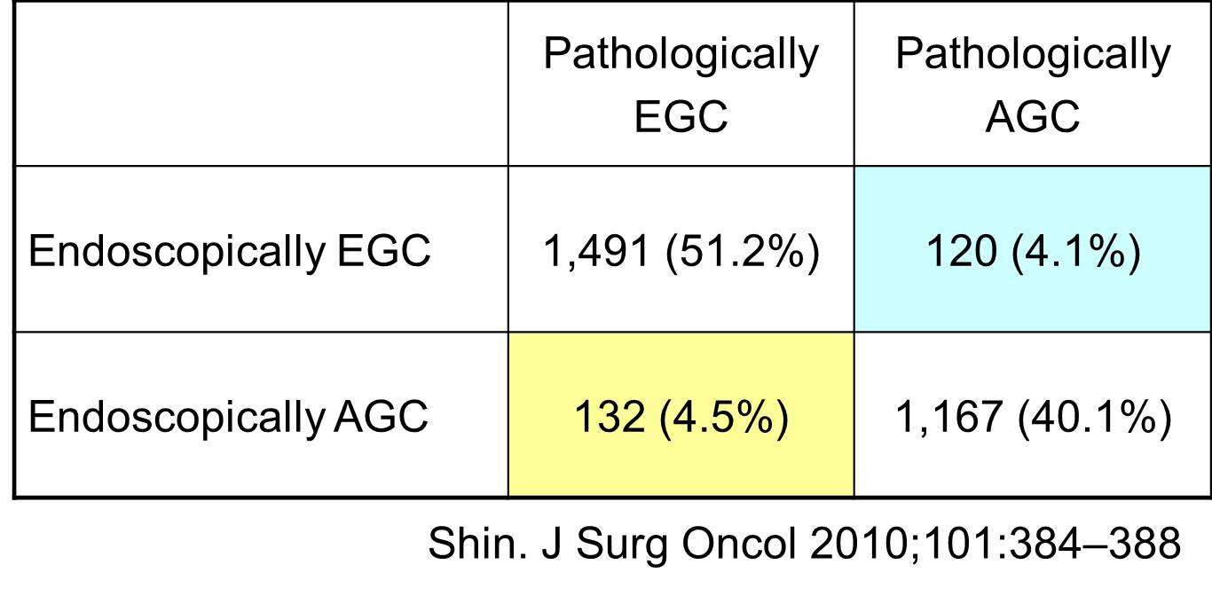
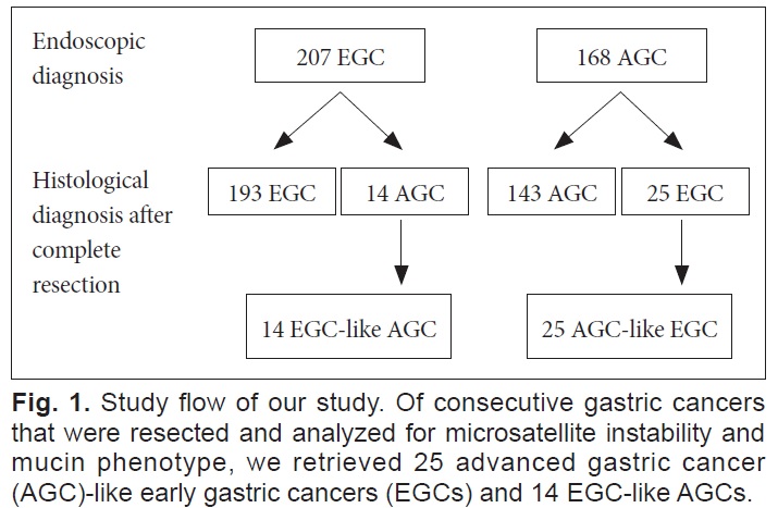
 [2014-11-2. 추가] Clinical Endoscopy 2013년 3월호에 AGC-like EGC, EGC-like AGC에 대한 건국대학교 이선영 교수님 팀의 논문이 있었습니다 (link). 결론을 인용합니다. Proximal part에 EGC-like AGC가 많다는 점에 동의합니다.
AGC-like EGCs predominate in the distal part of the stomach, while EGC-like AGCs predominate in the proximal part. When evaluating the depth of a gastric cancer, care should be taken not to underestimate measurements in proximal gastric cancers since they tend to be poorly-differentiated adenocarcinomas, in Lauren’s diffuse type, and invade deeper than their endoscopic appearance might suggest.
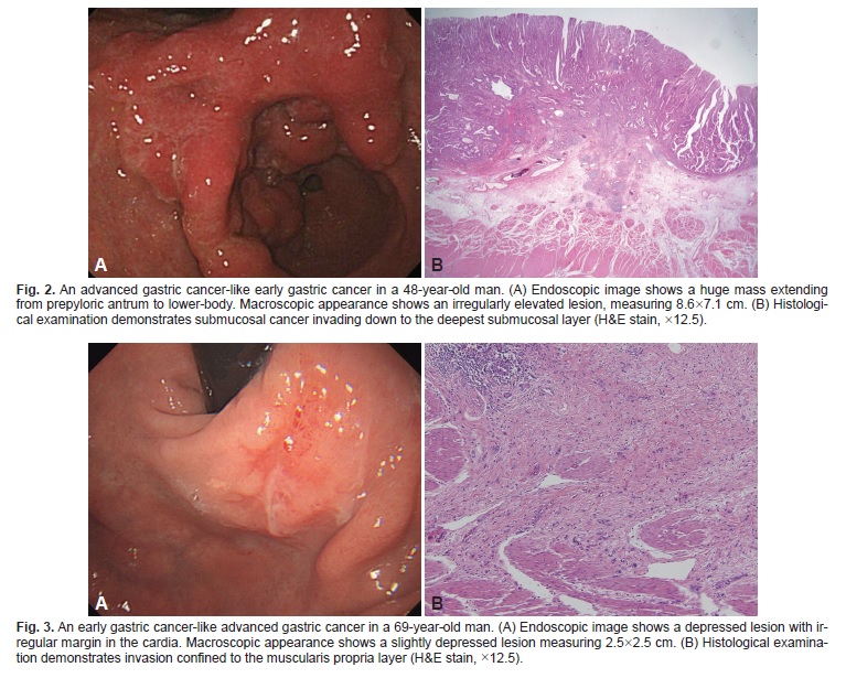
EGC와 AGC를 보다 정확히 구분하기 위하여 EUS를 하면 도움이 될까요? 저는 아니라고 믿습니다. 이 분야에 관심이 있어 많은 문헌들을 찾아보았지만 저의 믿음을 뒤집을 수 있을 데이터를 본 적이 없습니다.
[Related cases (EGC-like AGCs)]
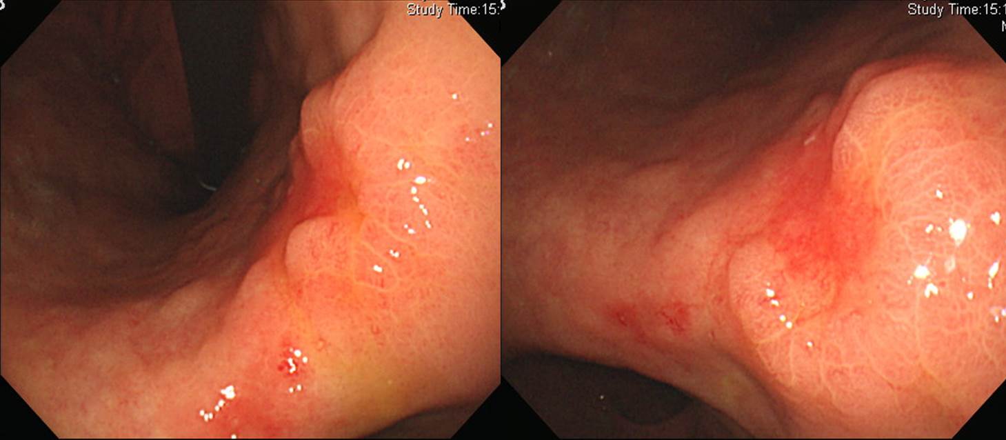
1. Location : middle third, center at body and posterior wall
2. Gross type : mimicking EGC type IIc
3. Histologic type : tubular adenocarcinoma, poorly differentiated
4. Histologic type by Lauren : diffuse
5. Size : 5x4x0.5 cm
6. Depth of invasion : extension to subserosa
7. Resection margin: free from carcinoma: safety margin
8. Lymph node metastasis : metastasis to 2 out of 35 regional lymph nodes (pN1)
9. Lymphatic invasion : present
10. Venous invasion : not identified
11. Perineural invasion : not identified
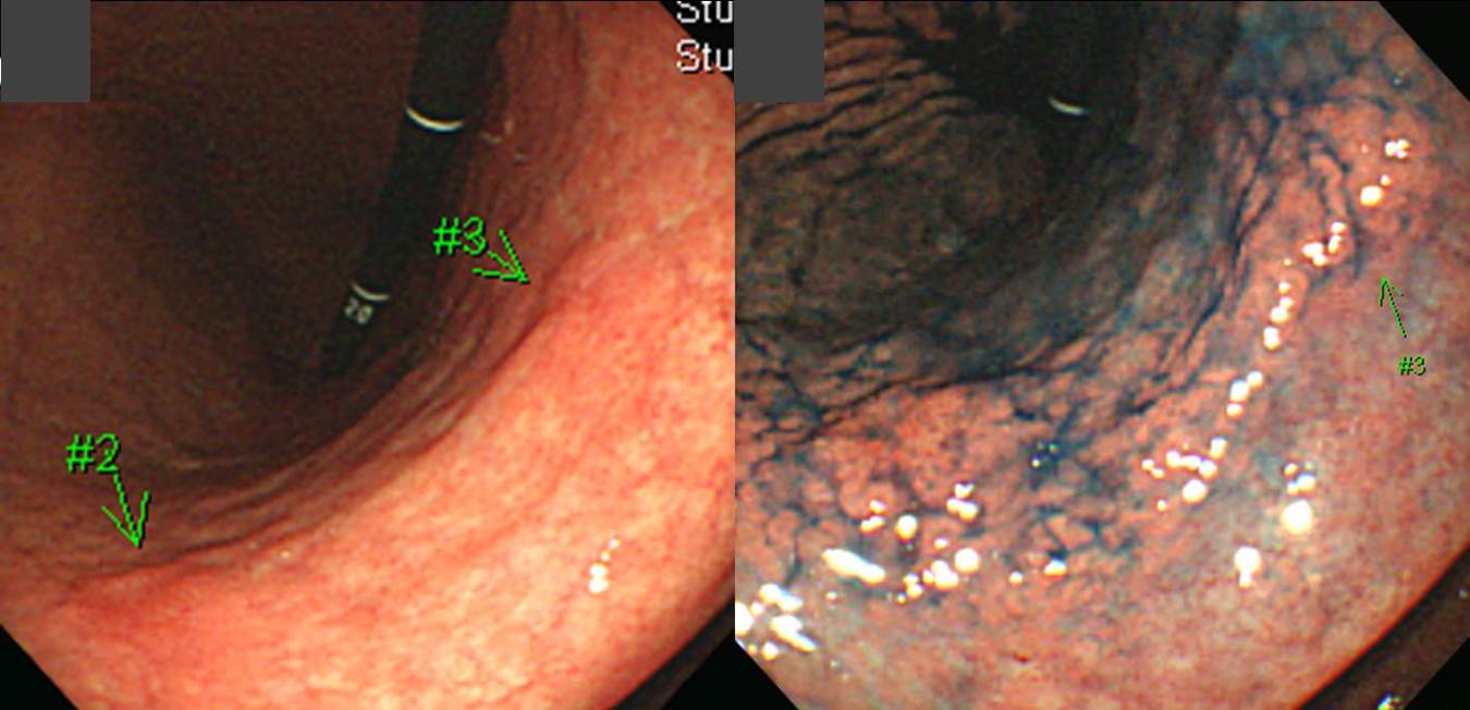
1. Location : middle third, center at body and lesser curvature
2. Gross type : Borrmann type (mimicking EGC type IIc+III)
3. Histologic type : signet-ring cell carcinoma
4. Histologic type by Lauren : diffuse
5. Size : 9.5x5.3x0.4 cm
6. Depth of invasion : extension to proper muscle (pT2a)
7. Resection margin: free from carcinoma: safety margin
8. Lymph node metastasis : metastasis to 9 out of 62 regional lymph nodes
9. Lymphatic invasion : present
10.Venous invasion : not identified
11.Perineural invasion : not identified
12.Associated findings : ulceration(ul IV)
© 2014.10.30. 이준행