EndoTODAY 내시경 교실
Beginner | ESA | Schedule | OPD
Seminars | Atlas | Recent | Links
![]() [Gastric cancer 633 - LC side of antrum can be a blind area]
[Gastric cancer 633 - LC side of antrum can be a blind area]
001 | 101 | 201 | 301 | 401 | 501 | 601 | 701 | 801 | 901 | 1000
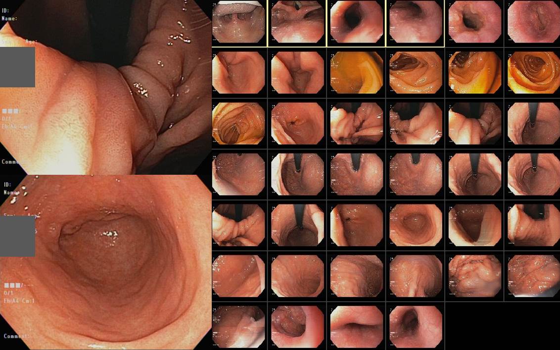
LC side of antrum can be a blind area. Surgery for EGC was done and the final pathology was mucosal cancer. I tried to find precancerous lesion in the previous endoscopy 2 years ago, but there was no picture of this area. There were 40 pictures for the endoscopy but lesser curvature side of the antrum cannot be documented.
위암 수술 환자입니다.

Stomach, subtotal gastrectomy: Early gastric carcinoma
1. Location : lower third Center at antrum and lesser curvature
2. Gross type : EGC type IIb
3. Histologic type : tubular adenocarcinoma, moderately differentiated
4. Histologic type by Lauren : mixed
5. Size : 3.4x3.1 cm
6. Depth of invasion : invades mucosa (muscularis mucosae) (pT1a)
7. Resection margin: free from carcinoma, safety margin: proximal 4.6 cm, distal 3.7 cm
8. Lymph node metastasis : no metastasis in 18 regional lymph nodes (pN0) (0/18: "3", 0/1; "4", 0/2; "5", 0/2; "6", 0/1; "7", 0/1; "9", 0/5; "8a", 0/3; "11p", 0/1; "12a", 0/2; "4sb", 0/0; "1", 0/0)
9. Lymphatic invasion : not identified
10. Venous invasion : not identified
11. Perineural invasion : not identified
12. AJCC stage by 8th edition: pT1a N0
정기적인 내시경 검사를 받던 분인지라 2년 전 내시경 사진에서 그 부위가 어떻게 보였는지 알아보고 싶었습니다. 아무리 자세히 살펴보아도 꼭 보고 싶은 바로 그 자리 사진이 없었습니다. 한 장은 병소 바로 위 angle, 다른 한 장은 병소 바로 아래 distal antrum이었습니다.
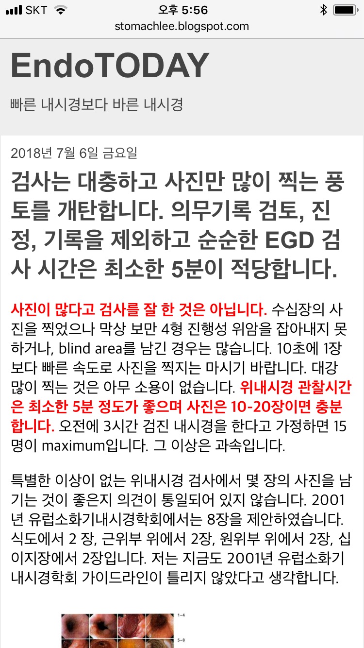
내시경 사진이 많다고 위 점막이 다 찍혀있을 것으로 생각하면 오해입니다. 이 환자의 경우 검사시간 196초 동안 40장의 사진이 있었습니다. 4.9초에 한 장인 셈입니다. 위내시경에서는 아주 많은 blind area가 있습니다. 좋은 습관을 가지는 것만이 blind area를 피하는 비결입니다.
이 환자에 해당하는 것은 아니지만... 검사는 대충하고 사진만 많이 찍는 풍토를 개탄합니다.
Blind area에서 발견된 위암입니다. 마지막 내시경은 39일 전이었습니다.
blind area에 발견된 진행성 위암
© 일원내시경교실 바른내시경연구소 이준행. EndoTODAY Endoscopy Learning Center. Lee Jun Haeng.