 EndoTODAY 내시경 교실
Beginner | ESA | Schedule | OPD
Seminars | Atlas | Recent | Links
![]() [Gastric cancer 691 - HNPCC 환자에서 우연히 발견된 위암]
[Gastric cancer 691 - HNPCC 환자에서 우연히 발견된 위암]
001 | 101 | 201 | 301 | 401 | 501 | 601 | 701 | 801 | 901 | 1000
10여년 전 대장암으로 수술을 받으신 HNPCC 환자가 위암이 발견되어 의뢰되었습니다. 일반적인 ESD 적응증에 해당하여 시술을 하였고 아래와 같은 결과였습니다.
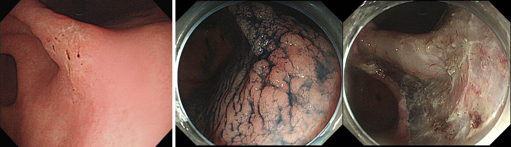
ESD: Early gastric carcinoma ;
1. Location : angle, posterior wall
2. Gross type : EGC type IIc
3. Histologic type : tubular adenocarcinoma, moderately differentiated
4. Histologic type by Lauren : intestinal
5. Size of carcinoma : (1) longest diameter, 18 mm (2) vertical diameter, 12 mm
6. Depth of invasion : invades mucosa (muscularis mucosa) (pT1a)
7. Resection margin : free from carcinoma(N), safety margin : distal 12 mm, proximal 11 mm, anterior 14 mm, posterior 12 mm, deep 600 ㎛
8. Lymphatic invasion : not identified(N)
9. Venous invasion : not identified(N)
10. Perineural invasion : not identified(N)
11. Microscopic ulcer : absent
12. Histologic heterogeneity: absent
일반적인 HNPCC surveillance를 하시도록 권하였고, 위암 ESD 후 추적관찰은 다른 환자와 비슷하게 하려고 합니다.
* 참고: EndoTODAY HNPCC
© 일원내시경교실 바른내시경연구소 이준행. EndoTODAY Endoscopy Learning Center. Lee Jun Haeng. (2018-11-9)