 EndoTODAY 내시경 교실
Beginner | ESA | Schedule | OPD
Seminars | Atlas | Recent | Links
![]() [Gastric cancer 742 - Incidentally found lung cancer during follow up after ESD for EGC]
[Gastric cancer 742 - Incidentally found lung cancer during follow up after ESD for EGC]
001 | 101 | 201 | 301 | 401 | 501 | 601 | 701 | 801 | 901 | 1000
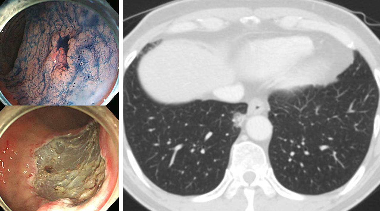
Lower lung field is covered by abdominal CT. A few years after a successful ESD for EGC, follow up abdominal CT was done. A small lung mass was found. Surgical consultation was done for r/o lung cancer.
Stomach, endoscopic submucosal dissection: Early gastric carcinoma
1. Location : antrum, posterior wall
2. Gross type : EGC type IIc
3. Histologic type : tubular adenocarcinoma, moderately differentiated
4. Histologic type by Lauren : intestinal
5. Size of carcinoma : (1) longest diameter, 8 mm (2) vertical diameter, 9 mm
6. Depth of invasion : invades mucosa (lamina propria) (pT1a)
7. Resection margin : free from carcinoma(N), safety margin : distal 10 mm, proximal 9 mm, anterior 18 mm, posterior 4 mm
8. Lymphatic invasion : not identified(N)
9. Venous invasion : not identified(N)
10. Perineural invasion : not identified(N)
11. Microscopic ulcer : absent
12. Histologic heterogeneity: absentLung and lymph node, right lower lobe, video assisted thoracotomy's lobectomy after wedge resection and mediastinal lymph node dissection: Adenocarcinoma, moderately differentiated, acinar pattern, medial basal segment:
(1) Size: 1.9x1.8x1.8 cm
(2) Vascular invasion (arteriolar or venous): not identified
(3) Lymphatic invasion: not identified
(4) Perineural invasion: not identified
(5) Negative resection margins (4.8 cm apart from main tumor)
(6) Pleural/extrapleural tumor superficially invading in the pleural connective tissue, but not beyond the elastic layer of the visceral pleura (PL0)
(7) Others
a. central scar
b. tumor-associated fibrosis (TAF)
c. micropapillary pattern, focal
(8) Regional lymph nodes included in main specimen (N1)
a. Total number examined: 0
b. Number involved by tumor: 0
(9) Separately submitted N1 or N2 lymph nodes
a. Total number examined: 21
b. Number involved by tumor: 0 (0/21: "7(Subcarinal)", 0/3; "10R(Hilar right)", 0/0; "11R(Interlobar right)", 0/1; "2R(Upper Paratracheal right)", 0/2; "4R(Lower Paratracheal right)", 0/3; "9R(Pulmonary ligament right)", 0/1; "2R-2", 0/2; "4R-2", 0/2; "7-2", 0/1; "7-3", 0/3; "11R-2", 0/1; "11R-3", 0/1; "11R-4", 0/1)
(10) TNM stage: pT1a N0
Additional pathologic findings
. Background lung : unremarkable
© 일원내시경교실 바른내시경연구소 이준행. EndoTODAY Endoscopy Learning Center. Lee Jun Haeng. (2019-4-18)