 EndoTODAY 내시경 교실
Beginner | ESA | Schedule | OPD
Seminars | Atlas | Recent | Links
![]() [Terminology. 내시경 용어] - 終
[Terminology. 내시경 용어] - 終
'EndoTODAY 내시경 삽입과 관찰'의 86쪽을 참고하시기 바랍니다.

![]() 1. 들어가는 말
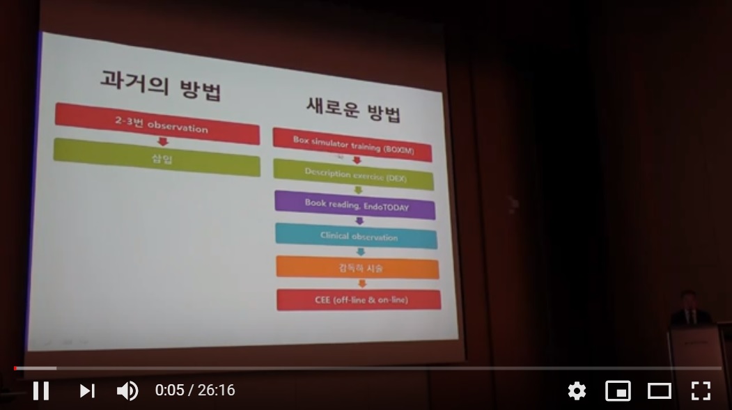
1. 들어가는 말
내시경 교과서에는 각 질병에서 관찰되는 소견이 자세히 씌여 있습니다. 그러나 소견을 기술하는 용어 자체에 대한 상세한 설명은 많지 않습니다. 본 강좌에서는 (1) 개념 혹은 정의가 명확하지 않은 용어를 내시경 의사의 입장에서 설명하고, (2) 내시경 의사마다 다르게 사용하는 흔한 용어를 증례 중심으로 검토하고자 합니다.
이렇게 자세히 기술하려면 내시경 용어의 뜻을 정확히 알아야 합니다.
내시경을 처음 배우는 분들이 겪는 어려움 중 하나는 적절한 용어의 선택입니다. 한 단어는 여러 의미를 가질 수 있습니다. 전문 영역에서는 일반적인 단어라고 하더라도 어떠한 context 혹은 뉘앙스가 추가되어 상식적인 의미와 제법 다르게 씌일 수 있습니다. 모든 전문 용어가 다 그렇습니다. 위내시경 영역도 마찬가지입니다. 내시경 용어는 민중서림 영어 사전을 찾아본다고 해결할 수 없는 어떠한 전문적인 뜻, 혹은 관행이 있습니다.
몇 개의 예 입니다. 'Mass'는 크기와 상관 없는 덩어리라는 뜻이지만, 내시경 영역에서는 진행성 암이 추정될 때 사용하고 있습니다. 조기 위암에 대하여 mass라고 쓰면 다들 놀랍니다. Ulcer의 edge와 margin도 마찬가지입니다. Edge는 선을, margin을 면을 나타나는 의미로 쓰이고 있습니다.
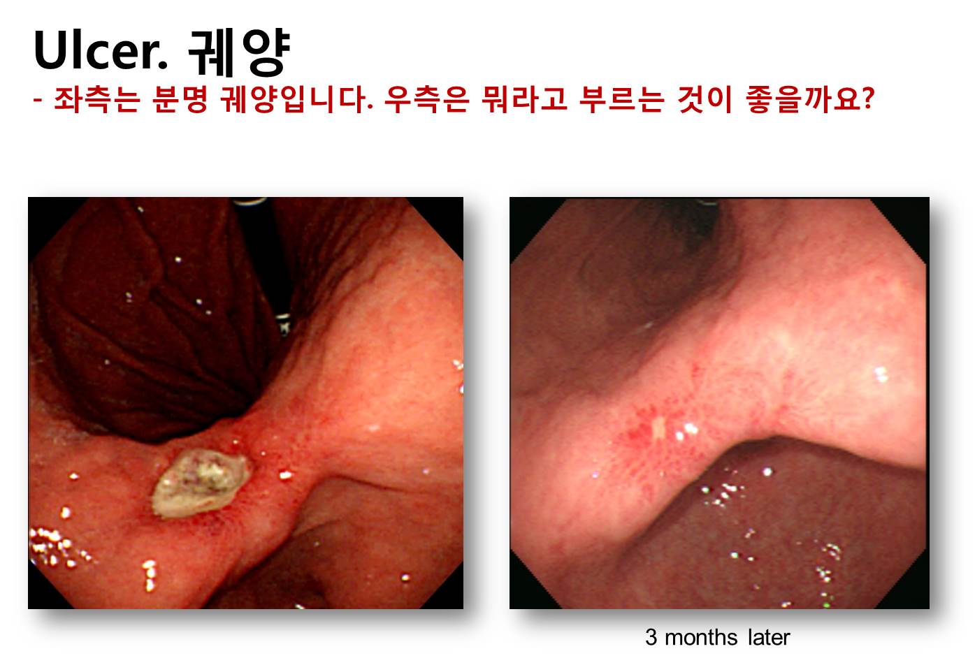
조직검사의 대원칙은 함몰형 병소의 edge에서 시행한다는 것입니다. 그런데 edge와 비슷한 용어로 margin이 있습니다. 이 둘은 매우 혼동되는 용어입니다. 사전을 찾아보다도 정확한 의미 차이를 알기 어렵습니다. 출판계에서는 edge와 margin을 정확하게 구분하여 사용하고 있습니다. 왼쪽 그림을 보시기 바랍니다. edge는 선이고 margin은 면입니다. 이와 같은 edge와 margin의 의미 차이를 고려하면 오른쪽 사진은 ‘궤양의 edge는 sharp하고 margin은 edematous하다’고 말할 수 있습니다. 이제 ‘조직검사는 edge에서 시행한다’는 것이 어디를 말하는지 정확히 이해하셨을 것입니다.
물론 '국제 표준 내시경 용어 사전'이라는 것이 없으므로 학파에 따라서 용어는 조금씩 다르게 사용될 수 있습니다. 일본 스타일과 우리나라 스타일은 제법 다릅니다. 여하튼 제가 말씀 드리는 것은 일원내시경교실에서는 mass, edge, margin이 그렇게 쓰인다는 것입니다. SMC style입니다.
![]() 2. 내시경 검사결과 용어 표준화의 필요성
2. 내시경 검사결과 용어 표준화의 필요성
내시경 검사결과 용어는 표준화되어야 합니다. 크게 두 가지 이유가 있습니다. 환자의 진료와 내시경 학습입니다. 내시경을 시행한 의사가 직접 검사 결과를 설명하고 치료까지 담당한다면, 검사결과 용어가 다소 표준을 벗어나도 큰 문제는 없습니다. 나중에 검사자 자신이 알아볼 수 있으면 그만이기 때문입니다. 만일의 사태(의료 분쟁 등)를 대비하여 제 3자가 철자법을 알아볼 수 있을 정도면 충분합니다.
내시경 검사를 시행한 의사와 환자를 진료하는 의사가 서로 다르다면 용어의 표준화는 매우 중요해집니다. 검사자와 치료자가 다른 경우는 매우 많으며 점차 증가하고 있습니다. (1) 개인병원이나 건진센터에서 내시경 검사를 받은 후 그 결과를 다른 의료기관에서 상담하는 경우, (2) 한 의료기관에서 치료를 받다가 추가 치료를 위하여 다른 의료기관으로 의뢰된 경우, (3) 내시경 검사를 시행하지 않는 의사가 내시경 처방을 내리는 경우, (4) 의무기록을 바탕으로 다른 의료기관에서 2차 의견을 구하는 경우 등입니다. 이와 같은 상황에서는 표준 용어를 이용하여 상세히 결과를 기술하고, 이를 뒷받침할 수 있는 질 좋은 사진을 남겨야 합니다. 내시경을 배우는 과정에서도 용어의 표준화는 중요합니다. 사실 내시경으로 관찰되는 현상을 어떻게 부를지 배우는 것이 내시경 교육의 핵심입니다.
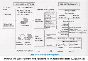
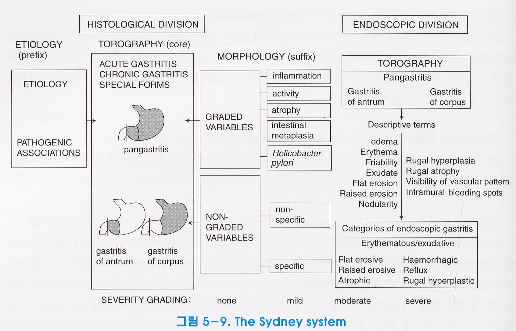
예를 들어, 만성 위염의 Sydney 분류는 부종 (edema), 홍반 (erythema), 부서지기 쉬움 (friability) , 삼출물 (exudate), 편평 미란 (flat erosion), 융기형 미란 (raised erosion), 결절성 (nodularity), 주름의 비대 (rugal hyperplasia), 주름의 위축 (rugal atrophy), 혈관 투영상 (visibility of vascular pattern), 내출혈반 (intramural bleeding spot) 등의 소견을 바탕으로 만성 위염을 7가지로 나누고 있습니다. 내시경에서 보이는 소견을 어떻게 부를지 명확하지 않다면 Sydney 방식에 따른 적절한 분류는 어려울 것입니다.
함몰형 조기위암과 양성 위궤양의 감별진단도 마찬가지입니다. 조기위암의 특징이 무엇인지 글로 쓰는 것은 비교적 쉽습니다. 그러나 내시경으로 관찰되는 소견을 적절히 이름짓지 못한다면 발견한 병소가 조기위암인지 알 수 없을 것입니다. 검사자가 사용하는 용어가 부적절하다면 교과서적 지식이 많더라도 정확한 진단은 불가능합니다.
![]() 3. 내시경 용어는 대부분 피부과 용어를 차용한 것입니다.
3. 내시경 용어는 대부분 피부과 용어를 차용한 것입니다.
피부과 의사는 병변을 보고 진단을 붙입니다. 내시경 의사도 병변을 보고 진단을 붙입니다. 직접 보느냐, 기구를 통하여 보느냐의 차이가 전부입니다. 보이는 것을 표준 용어로 기술하고 적당한 진단을 찾는 과정은 동일합니다. 피부의 병변은 고대부터 기술되어 왔습니다. 비교적 명확히 정의되어 있습니다. 궤양 (ulcer), 미란 (erosion), 발적 (hyperemia), 반점 (patch), 삼출물 (exudate), 과형성 (hyperplasia), 비후 (hypertrophy), 부종 (edema), 용종 (polyp), 종괴 (mass) 등 내시경 용어의 뿌리는 피부과입니다. 용어 정의가 없는 내시경 교과서가 많습니다. 내시경 교과서의 설명이 부족한 경우 피부과 의사의 자문을 구하거나 피부과 교과서를 참조하면 도움이 됩니다.
![]() 4. 융기형 병소를 표현하는 용어
4. 융기형 병소를 표현하는 용어
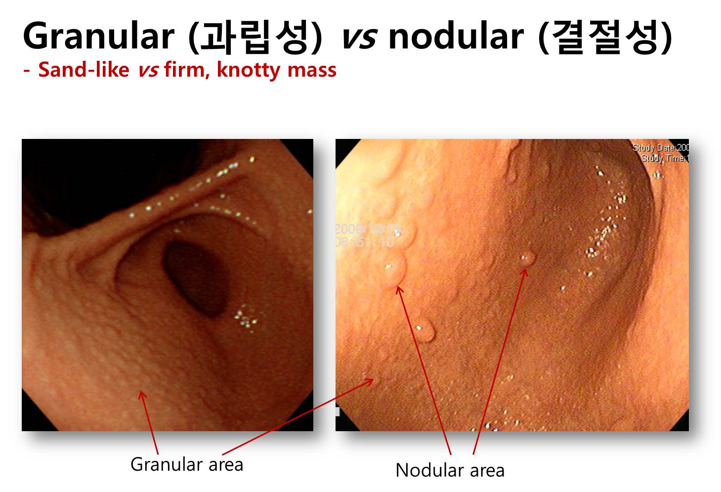
점막의 넓고 융기된 표면을 표현하는 용어는 과립성 (granular), 결절성 (nodular), 조약돌 (cobble-stone) 등입니다. 과립성이 가장 작고, 조약돌이 가장 큽니다.
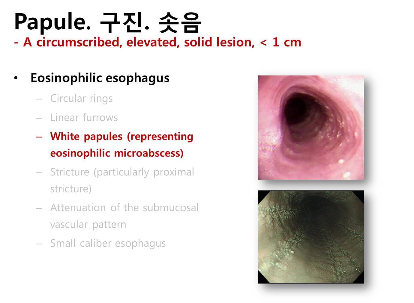
국소 융기병소를 표현하는 용어는 구진 (papule), 판 (plaque), 결절 (nodule), 종양 (tumor) 등입니다. 구진과 판은 염증성 병소를 표현하는데 사용되고, 결절과 종양은 종양성 병소를 표현하는데 사용됩니다. Tumor island는 궤양형 위암 바닥의 돌출부를 말합니다.
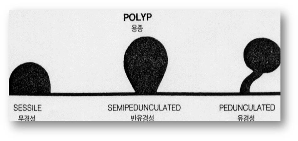
용종을 표현하는 방법은 다양합니다. 가장 전통적인 방법은 무경성 (sessile), 반유경성 (semipedunculated), 유경성 (peduculated)입니다. 최근에는 Kudo 방식이 보편적이지만 과거에는 Yamada 분류도 널리 사용되었습니다. 곰팡이모양 (fungating)은 매우 지저분하고 빠르게 자라는 종양을 표현하는 피부과 용어입니다. 위장관 내시경에서는 가급적 사용하지 않는 것이 좋을 것 같습니다.
![]() 5. 함몰형 병소를 표현하는 용어
5. 함몰형 병소를 표현하는 용어
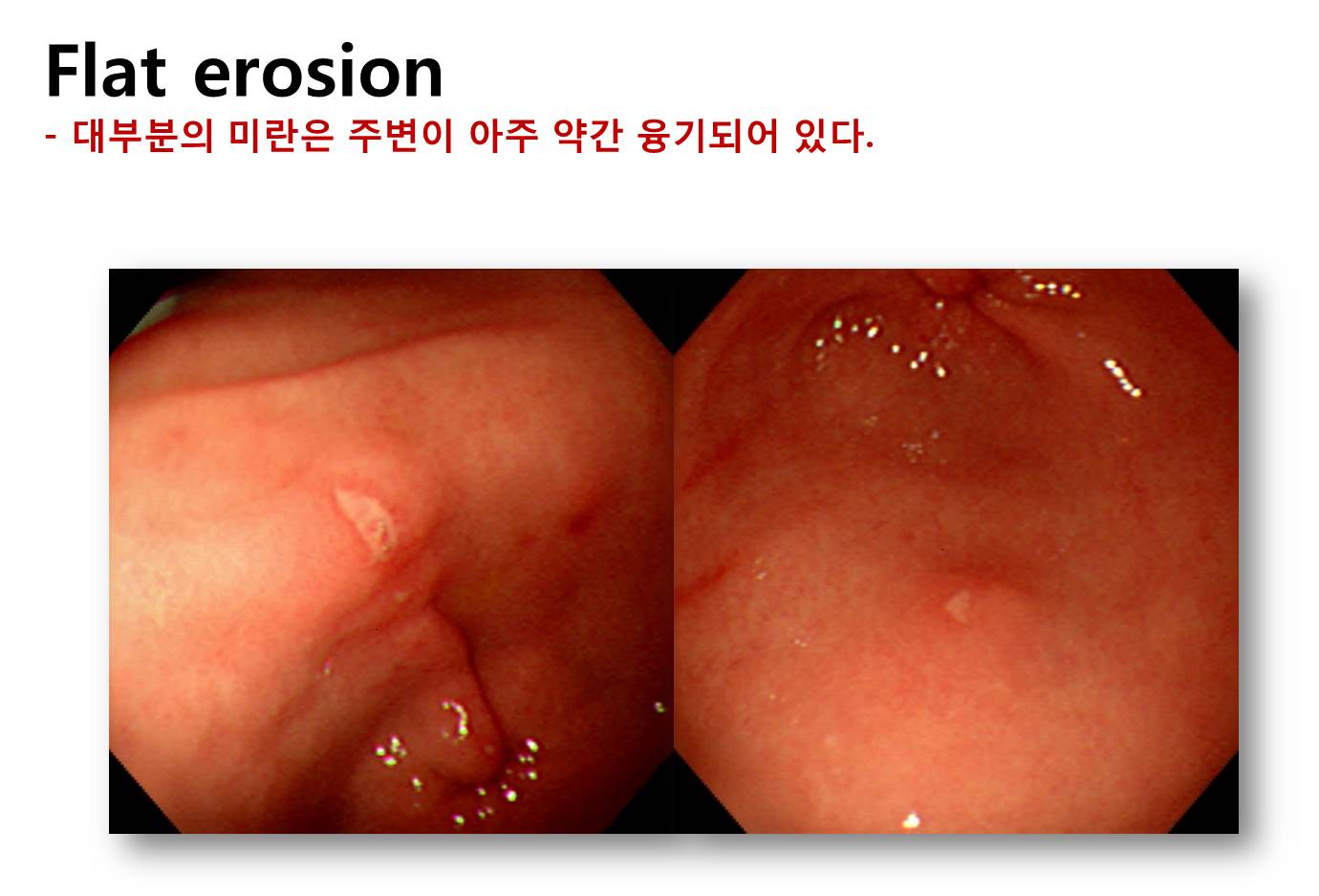
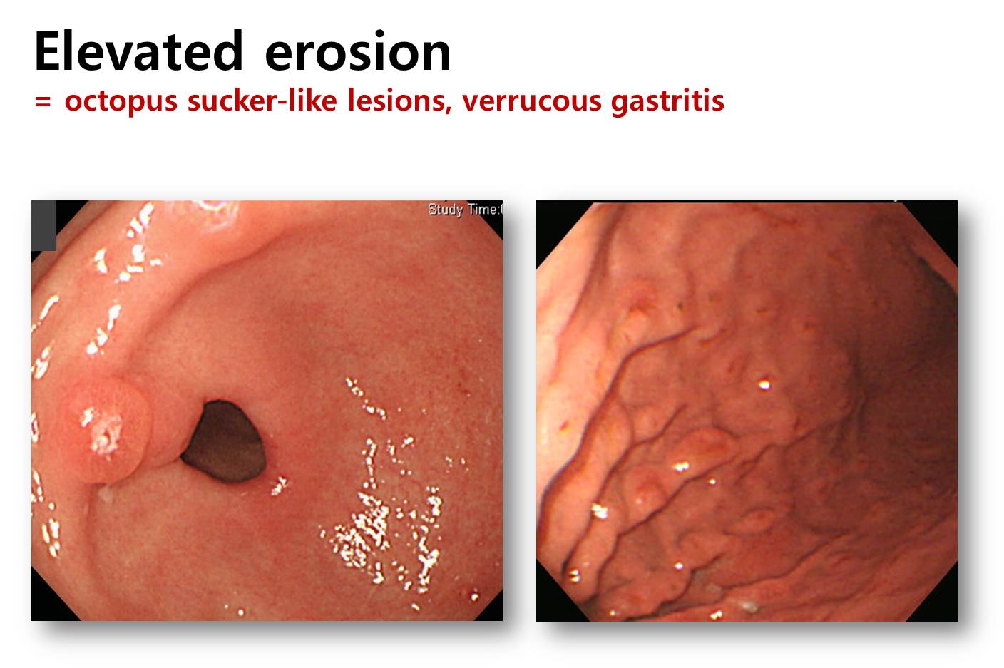
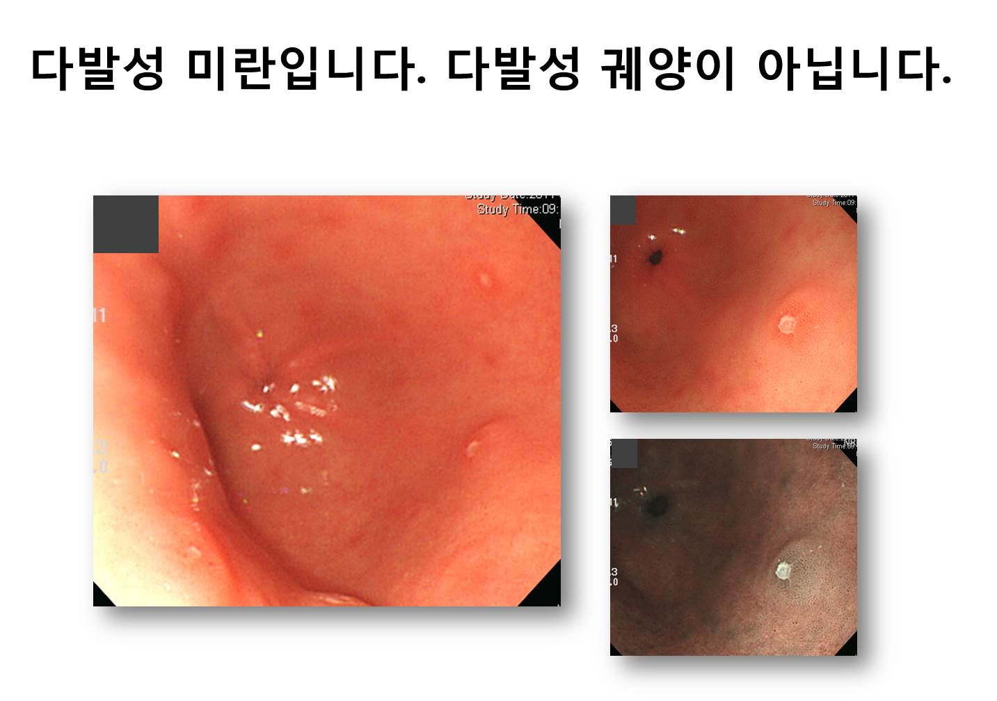
대표적인 함몰형 병소는 미란 (erosion)과 궤양 (ulcer)입니다. 병리학적으로 점막층에 국한된 조직 결손을 미란, 점막하층 이하까지의 조직 결손을 궤양이라고 합니다. 내시경으로 미란과 궤양을 정확히 구분하기는 어렵습니다. 뚜렷한 함몰부를 가진 비교적 큰 병소는 궤양으로, 작고 얕은 병소는 미란으로 부르는 것이 일반적입니다. 위내시경에서 궤양인지 미란인지 명확하지 않은 경우 미란으로 부를 것을 권하고 싶습니다. 미란을 궤양으로 부르면 여러 불필요한 일이 벌어지기 때문입니다. 조직검사가 필요하고, 위암의 위험성을 경고해야 하고, 헬리코박터 검사 및 치료가 필수적이고, 추적관찰이 필요하고, 비스테로이드소염진통제를 사용할 때 프로톤펌프억제제를 함께 처방해야 하는 등 미란에서는 불필요한 많은 일들이 궤양에서는 필요합니다. 아프타 (aphtha)는 붉은 띠를 가진 5 mm 이하의 작고 얕은 함몰병소입니다.
![]() 6. 동양식 접근법과 서양식 접근법
6. 동양식 접근법과 서양식 접근법
우리 나라의 내시경은 일본을 통하여 도입되었습니다. 아직도 일본 내시경책이 번역되어 널리 읽히고 있습니다. 일본과 서양의 학문하는 태도는 크게 다릅니다. 일본은 전통을 따르는 경향이고, 서양은 정의나 객관성을 중요시 합니다. 우리는 우리 나름의 방식이 있어야 하는데, 조기위암 내시경 진단 부분에서는 아직 일본의 영향이 큰 상태입니다.
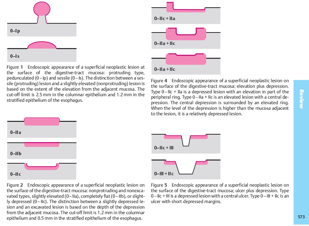
일본에서 IIc형 조기위암은 “주위 점막보다 약간 함몰된 조기위암”으로 정의됩니다. 그러나 주름이 끌려온 IIc형 조기위암, 특히 주름의 일부까지 암이 침윤된 경우에는 주위 점막보다 함몰되지 않은 경우가 있습니다. 편평하거나 오히려 약간 융기되기까지 합니다. 관례에 따라서 어떤 모양은 함몰형 조기위암으로 부르는 것이 당연시될 뿐, 실제 개별 병소가 함몰되어 있는지 아닌지는 무시됩니다. IIc형 조기위암과 III형 조기위암의 구분, I형 조기위암과 IIa형 조기위암의 구분에서 이러한 경향은 더욱 두드러집니다. 일본에서는 약간 올라와 있으면 IIa형, 좀 더 올라와 있으면 I형입니다. 그 둘의 구분은 명확히 정의되지 않았습니다. 많은 증례를 보면서 서로 눈높이를 맞추었기 때문에 일본 내시경 의사들의 분류법은 상당히 일관성이 있습니다. 장기간의 도제교육 영향이라고 생각됩니다.
서양사람들은 이와 같은 일본식 분류를 이해하지 못합니다. 조기위암 내시경 분류를 배우는 서양 사람들은 자꾸 묻습니다. “I형과 IIa을 나누는 기준은 무엇입니까?” 혹은 “IIc형과 III형을 나누는 기준은 무엇입니까?” 일본사람들이 답합니다. “이 사진과 같은 것이 I형이고 저 사진과 같은 것이 IIa형입니다. 많은 사진을 보면 저절로 알게 됩니다.” 서양사람이 또 묻습니다. “그래도 모르겠습니다. 좀더 객관적인 기준은 없습니까?” 이러한 질의응답 끝에 탄생한 것이 Paris 분류입니다. Paris 분류에 따르면 I형과 IIa형을 나누는 기준은 2.5mm, IIc형과 III형을 나누는 기준은 1.2mm입니다. 2.5mm나 1.2mm를 정확히 알기 어렵기 때문에 조직겸자의 두께와 비교하라는 권유도 있습니다.
수 십 년 동안 별 불편함이 없이 사용되던 조기위암 내시경 분류에 뜸금없이 밀리미터 단위의 기준이 들어간 것은 서양 사고방식의 영향입니다. 내시경 전문가들에게 물어보면 Paris 분류에서 제시된 밀리미터 기준을 거의 사용하지 않는다고 합니다. 편안하게 사용하던 과거의 분류법에 억지 기준을 추가함으로써 일만 복잡해진 경우입니다.
설명: 그림은 근사하지만 실제 분류에서 크게 도움이 되지 않습니다. 일본 방식을 서양 사람에게 설명하기 위한 고육지책 정도로 생각하면 좋겠습니다.
설명: 아무도 2.5mm를 고려하여 융기형 조기위암을 분류하지 않습니다.
설명: 아무도 1.2mm를 고려하여 함몰형 조기위암을 분류하지 않습니다.
![]() 7. 논문에서 사용되는 용어
7. 논문에서 사용되는 용어
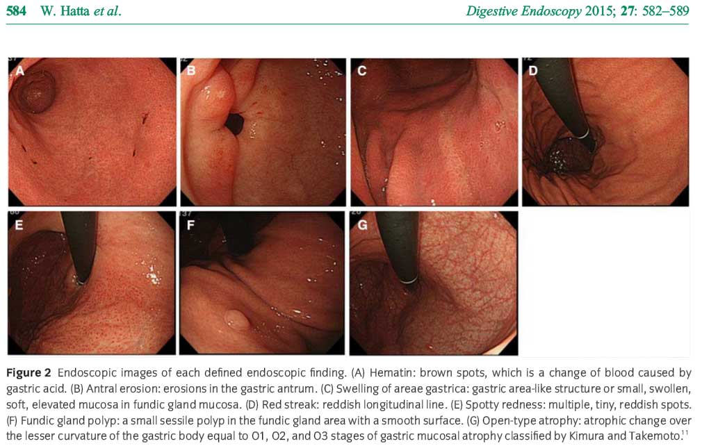
위내시경 소견으로 위산 분비를 어느 정도 예측할 수 있다는 흥미로운 일본 논문을 소개합니다 (Hatta. Digest Endosc 2015). Hematin과 antral erosion은 위산이 많을 때, area gastritis swelling과 open type atrophy는 위산이 적을 때 보이는 소견이라고 합니다. 연구자가 내시경 소견을 어떻게 불렀는지 용어 사용법을 참고하시기 바랍니다. 제게는 (c) swelling of area gastrica와 (e) spotty redness의 구분이 어려웠습니다. (2015-8-8)
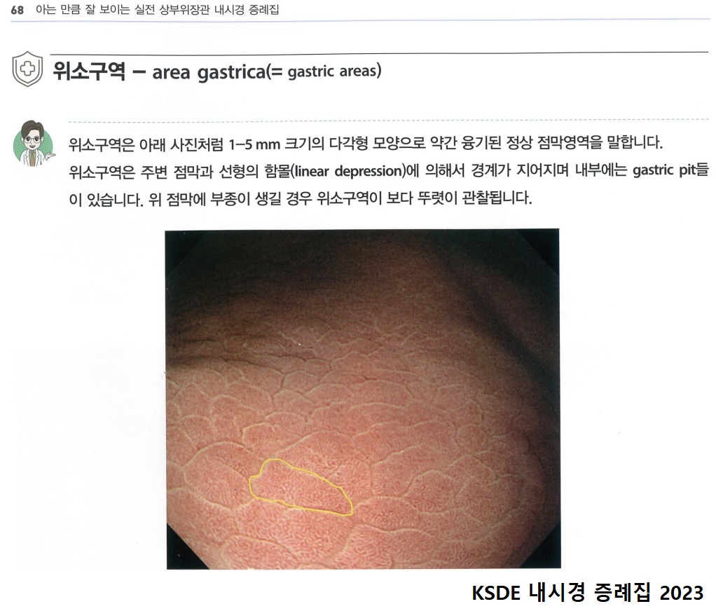
어떤 책에서 area gastrica 위소구를 설명한 부분을 옮깁니다.
![]() 8. 내시경 결과를 어떻게 쓸 것인가? (2020-2-16. 이준행)
8. 내시경 결과를 어떻게 쓸 것인가? (2020-2-16. 이준행)
내시경 검사에서 발견된 결과를 어떻게 쓸 것인가에 대한 표준안은 없습니다. 다만 위암 환자의 내시경 결과에는 병소의 위치, 크기, 모양 이외에 정부에서 정한 적정성지표 기준이 충족되어야 할 것 같습니다. 다소 어이없지만 칼자루를 쥐고 있는 쪽에서 정한 것이니 어쩔 수 없이 따라야 합니다. 안타깝습니다.
흔히 누락되는 항목은 조기위암의 궤양 유무, 진행성 위암의 크기입니다. 과거와 같이 huge ulceroinfiltrative mass in the GC side of lower body라고 쓰고 예쁜 사진을 찍어두면 엉터리 내시경 검사로 간주됩니다. 크기가 빠졌기 때문입니다. 이게 말이 됩니까? 그래도 어쩔 수 없습니다. 따르는 수밖에...
2020년 현재 적용되고 있는 기준은 아래와 같습니다.
2019년 위암적정성 평가 기준
2020년 현재 적용되고 있는 기준에 따라 아래와 같은 지적이 있었습니다. 위암의 type이나 크기가 빠진 경우가 많다고 합니다.
잘 된 사례와 잘 못 된 사례
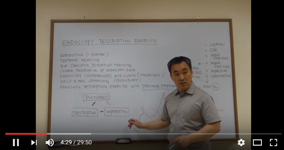
이래 저래 안전한 방법은 (1) 위치, (2) 크기, (3) 주소견, (4) 부소견, (5) 진단, (6) 분류 순서로 작성하는 것입니다. Description exercise workshop (DEX)에서 체계적인 내시경 소견 기술법을 배우실 수 있습니다.
* 참고: EndoTODAY Description exercises workshop
![]() 9. Skin lesions
9. Skin lesions
내시경 용어는 피부과 용어에서 차용한 것이 많습니다. 피부과 용어를 잘 알면 내시경 용어도 잘 사용할 수 있습니다.
Common skin lesions.
(1) Macules: These are areas of skin discoloration which are neither raised nor depressed.A macule is a flat, distinct, discolored area of skin less than 1 centimeter (cm) wide. A large macule i.e Areas of discoloration that are larger than 1 cm are referred to as patches.
Macules and Patches
(2) Papules: These are circumscribed elevations of skin which are palpable with no visible fluid and diameter is less than 5 mm or upto 10 mm in diameter at the widest point.
Papule and Plaque
(3) Plaque: It is actually a broad papule, or confluences of papules which is greater than 1 cm.
(4) Nodules: It is same as papules but the depth of nodules is more than 10 mm. Remember! it is the depth which differentiate nodules from papules.
Nodule
(5) Vesicles: These are cystic swellings containing serous fluid and diameter is up to 5 mm.
(6) Bullae: These are cystic lesions of more than 5 mm containing serous , seropurulent or hemorrhagic fluid.
Vesicles and bullae
(7) Postules: These are small elevation of the skin similar to vesicles containing opaque, cloudy or purulent material usually consisting of necrotic inflammatory cells. These can be either white or red.
(7) Wheals: These are swellings of skin due to acute localized edema. It usually disappear within 24 to 48 hours. The temporary raised bubble of taut skin on the site of a properly-delivered intradermal injection is also called a welt, with the ID injection process itself frequently referred to as simply “raising a wheal” in medical texts
Wheal
(8) Cyst: A cyst is an epithelial-lined cavity containing liquid, semi-solid, or solid material.
(9) Erosion: An erosion is a discontinuity of the skin exhibiting incomplete loss of the epidermis a lesion that is moist, circumscribed, and usually depressed.
(10) Ulcer: An ulcer is a discontinuity of the skin exhibiting complete loss of the epidermis and often portions of the dermis and even subcutaneous fat.
(11) Fissure: A fissure is a crack in the skin that is usually narrow but deep.
Comparison of skin lesions
(12) Telangiectasia: A telangiectasia represents an enlargement of superficial blood vessels to the point of being visible.
(13) Burrow: This appears as a slightly elevated, grayish, tortuous line in the skin, and is caused by burrowing organisms.
Secondary skin lesions:
(1) Scale: It is dry or greasy laminated masses of that represent thickened stratum corneum.
(2) Crust:It is a dried serum, pus, or blood usually mixed with epithelial and sometimes bacterial debris.
(3) Lichenification: The epidermal thickening characterized by visible and palpable thickening of the skin with accentuated skin markings.
(4) Excoriation: A punctate or linear abrasions produced by mechanical means (often scratching), usually involving only the epidermis, but commonly reaching the papillary dermis.
(5) Imduration: Dermal thickening causing the cutaneous surface to feel thicker and firmer.
(6) Atropy: It refers to a loss of tissue, and can be epidermal, dermal, or subcutaneous. With epidermal atrophy, the skin appears thin, translucent, and wrinkled.Dermal or subcutaneous atrophy is represented by depression of the skin.
(7) Maceration: It is softening and turning white of the skin due to being consistently wet.
(8) Umbilication: Formation of a depression at the top of a papule, vesicle, or pustule.
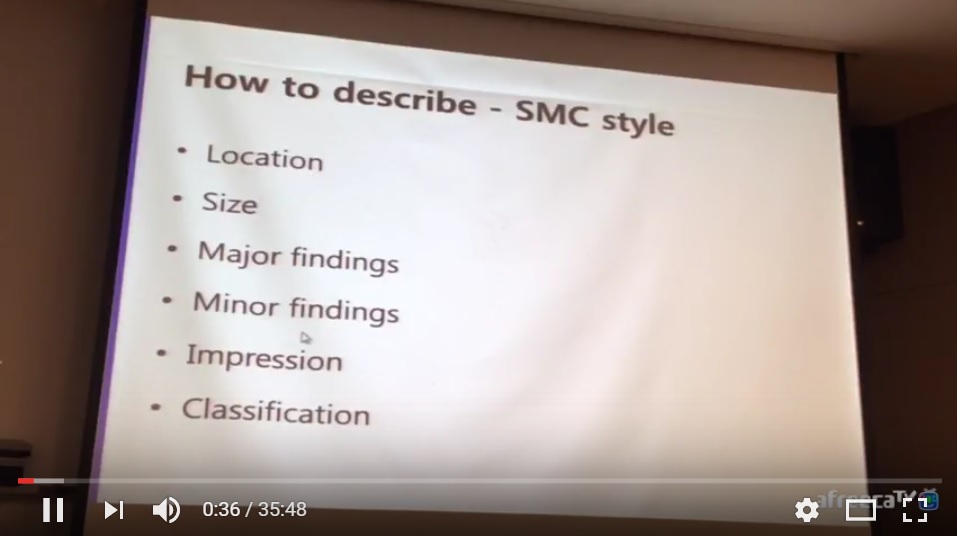
![]() 10. SMC description style
10. SMC description style
Follow the description style: (1) Location, (2) Size, (3) Major findings, (4) Minor findings, (5) Impression, (6) Classification
2017-7-31. 3년차 전공의 첫 내시경 description orientation
Description은 SMC style을 따라 주십시요.
내시경 description이 적절한지 살펴보는 기준(= 축, axis)은 두 가지 입니다.
몇 개의 증례를 바탕으로 SMC style로 description을 해 보겠습니다.
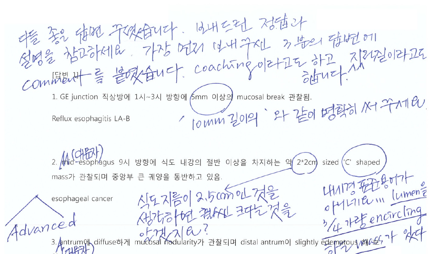
중부식도의 내강을 좁히는 5cm 크기의 protruded mass with central ulceration이 보입니다. 일부 표면은 약간 nodular하고 부분적인 friability가 있습니다. Advanced esophageal cancer, type 3
위전정부 소만에서 후벽쪽으로 3cm의 ulcer가 있습니다. Edge는 sharp하고 margin은 약간 edematous하며, 궤양 바닥은 비교적 균일하며 white exudate로 덮혀있습니다. 비정상적인 fold 변화는 없습니다. BGU A1
위체상부부터 근위전정부까지 약 15cm에 걸쳐 위벽이 두껍고 잘 펴지지 않는 소견입니다. Fold는 nodular하게 두꺼워져있고 fold 사이의 간격은 좁아져 있으며 일부 fold는 서로 융합되어 있습니다. 위체하부 대만의 두꺼워진 fold의 상단에는 작은 궤양 부위가 관찰됩니다. AGC Borrmann type 4
위체하부 대만에 약 1.5cm 크기의 depressed lesion이 관찰됩니다. Edge는 sharp하고 margin은 정상적이며 바닥은 비교적 flat하며 발적과 퇴색이 섞여 있습니다. Converging folds는 없으나 함몰병소로 자체로 인하여 정상 fold가 끊겨졌고, 함몰 병소 바로 옆을 지나가는 fold는 일부 함몰부위로 끌려오는 모양입니다. EGC IIc
위전정부에 2-4mm의 multiple pale slightly nodular lesion들이 비교적 균일하게 scattered 되어 있습니다. Metaplastic gastritis
과거에 어떤 분께서 제게 답변을 보내주셔서 제가 feedback 한 내용의 일부입니다. 이와 비슷한 방식으로 소견과 진단을 보내주면 됩니다.
[2016-2-14. 애독자 질문]
매일 좋은 강의 잘 보고 있습니다. 교수님이 강의해주신 내시경 용어에 대해 궁금한 점이 있어 메일 보냅니다
Granular와 nodular의 차이를 얘기할때 피부과에서는 크기의 기준이 1cm입니다. 그러나 내시경에서도 1cm을 기준으로 하시는지요?
공부를 하다보니 LST분류에서 granular type 중 nodular mixed type으로 진단하는 기준이 도움될 듯해서 찾아보았습니다. 파리분류에서는 IIa +Is 가 nodular mixed type으로 기술되어서 nodular 의 기준이 1cm보다는 작아보입니다. 높이 기준은 있지만 크기기준이 없어서 많이 혼동됩니다. 교수님은 어느 정도를 nodular로 진단하시는지요?
좋은 의견부탁드립니다. 대구에서 XXX 올림
[2016-2-14. 이준행 답변]
좋은 질문 감사합니다. 크기에 대한 정답은 없습니다. Granular는 sand-like이고 nodular는 knot-like입니다. 육안소견이 중요한 피부과에서 1 cm을 기준으로 나누는 것은 나름 타당합니다. 그러나 늘 확대된 영상을 바라보는 - 확대내시경이 아닌 보통 내시경도 확대된 영상입니다 - 내시경 의사 입장에서는 1 cm 기준은 너무 큰 듯 합니다.
정답은 아니더라도 우리끼리 대강의 기준을 만들면 좋을 것 같습니다. 그래서 저는 대강 5 mm 이상을 nodular로 부르도록 가르치고 있습니다.
쌀알 사진입니다. 쌀알의 장경은 대강 5 mm입니다. 쌀알보다 크면 nodular... 그래서 기준이 5 mm. 어떻습니까. 너무 작위적인가요? 이것 말고도 제 맘대로 정한게 한두가지가 아닙니다.

[2016-2-14.애독자 답변]
좋은 의견 감사합니다 저도 개인적으로 5mm를 기준으로 생각하고 있습니다. 위장에서 조기위암분류에서 Type 0-I과 type0-IIa 의 높이 기준이 2.5mm인것으로 되어있습니다. 이 기준을 적용하기에는 크기가 너무 작은 것 같고 피부과 기준대로 1cm은 너무 큰 것 같아서 저도 5mm가 가장 합당한 것으로 생각합니다.
![]() [References]
[References]
2) Dixon MF, Genta RM, Yardley JH, et al. Classification and grading of gastritis. The updated Sydney System. International Workshop on the Histopathology of Gastritis, Houston 1994. Am J Surg Pathol 1996;20:1161-81.
3) Kudo S. Endoscopic mucosal resection of flat and depressed types of early colorectal cancer. Endoscopy 1993;25:455-61.
4) Endoscopic Classification Review G. Update on the paris classification of superficial neoplastic lesions in the digestive tract. Endoscopy 2005;37:570-8.
5) Daily EndoTODAY 내시경 용어 (2012/5/7 - 2012/5/28)
6) Sun-Young Lee. Endoscopic gastritis, serum pepsinogen assay, and Helicobacter pylori infection Korean J Intern Med 2016
© 일원내시경교실 바른내시경연구소 이준행. EndoTODAY Endoscopy Learning Center. Lee Jun Haeng.


