 EndoTODAY 내시경 교실
Beginner | ESA | Schedule | OPD
Seminars | Atlas | Recent | Links
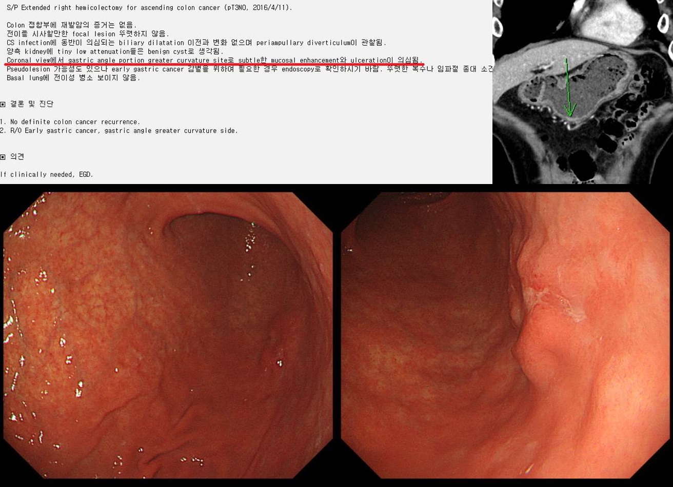
![]() [Gastric cancer 783. A small EGC incidentally found in abdominal CT after colon cancer surgery]
[Gastric cancer 783. A small EGC incidentally found in abdominal CT after colon cancer surgery]
001 | 101 | 201 | 301 | 401 | 501 | 601 | 701 | 801 | 901 | 1000
In clinical practice, CT is usually done after endoscopy. The EGC lesion can be found or suspected in CT based on endoscopic findings. Without prior endoscopic diagnosis, EGC is very difficult to find in CT.
In follow up abdominal CT after colon cancer surgery, a subtle mucosal enhancement and ulceration was found in CT. Endoscopy showed a flat elevated type EGC in that location. Surgery was done.
Stomach, radical subtotal gastrectomy: Early gastric carcinoma
1. Location : middle third, Center at lower body and greater curvature
2. Gross type : EGC type IIb+IIc
3. Histologic type : tubular adenocarcinoma, mixed moderately and poorly (solid) differentiated
4. Histologic type by Lauren : mixed
5. Size : 2.5x2.0 cm
6. Depth of invasion : invades submucosa (sm3) (pT1b)
7. Resection margin: free from carcinoma, safety margin: proximal 10.0 cm, distal 9.5 cm
8. Lymph node metastasis : no metastasis in 24 regional lymph nodes (pN0)
9. Lymphatic invasion : not identified
10. Venous invasion : not identified
11. Perineural invasion : not identified
12. Peritoneal cytology : negative
13. AJCC stage by 8th edition: pT1b N0
It is happy to work with an excellent radiologist.
© 일원내시경교실 바른내시경연구소 이준행. EndoTODAY Endoscopy Learning Center. Lee Jun Haeng. (2019-8-26)