EndoTODAY 내시경 교실
Beginner | ESA | Schedule | OPD
Seminars | Atlas | Recent | Links
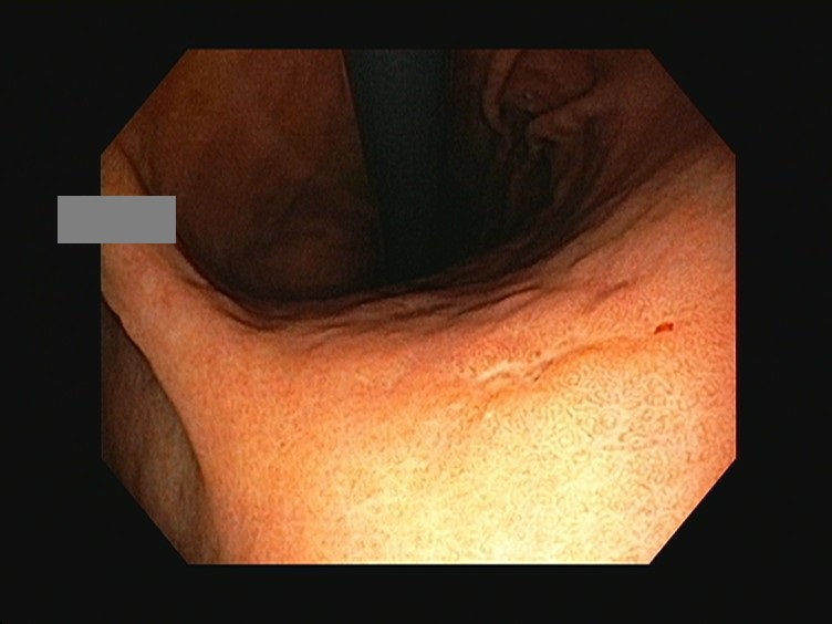
![]() [Gastric cancer 796. Biopsy was signet ring cell carcinoma and ESD pathology was mostly poorly differentiated adenocarcinoma]
[Gastric cancer 796. Biopsy was signet ring cell carcinoma and ESD pathology was mostly poorly differentiated adenocarcinoma]
001 | 101 | 201 | 301 | 401 | 501 | 601 | 701 | 801 | 901 | 1000
A small depressed lesion with uneven margin was found on the posterior wall of the lower body. Biopsy result was signet ring cell carcinoma. After referral, outside slide review was focal atypical cells, signet ring cell carcinoma. What would you do?
After careful discussion with the patient (M/40), I performed the ESD. Because the tumor border was unclear, I tried to secure enough cancer free margin.
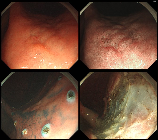
ESD: Early gastric carcinoma
1. Location : lowbody, posterior wall
2. Gross type : EGC type IIb+IIc
3. Histologic type : tubular adenocarcinoma, poorly differentiated, with signet ring cell carcinoma (20%)
4. Histologic type by Lauren : diffuse
5. Size of carcinoma : (1) longest diameter, 16 mm (2) vertical diameter, 12 mm
6. Depth of invasion : invades mucosa (muscularis mucosa) (pT1a)
7. Resection margin : free from carcinoma(N), safety margin : distal 8 mm, proximal 9 mm, anterior 10 mm, posterior 16 mm, deep 300
8. Lymphatic invasion : not identified(N)
9. Venous invasion : not identified(N)
10. Perineural invasion : not identified(N)
11. Pre-existing adenoma : none
12. Microscopic ulcer : absent
13. Histologic heterogeneity: absent
The intial forceps biopsy result was signet ring cell carcinoma, but the final ESD pathology was mostly poorly differentiated adenocarcinoma. I would follow up very carefully.
© 일원내시경교실 바른내시경연구소 이준행. EndoTODAY Endoscopy Learning Center. Lee Jun Haeng. (2019-9-14)