EndoTODAY 내시경 교실
Beginner | ESA | Schedule | OPD
Seminars | Atlas | Recent | Links
![]() [Gastric cancer 797. Is it an EGC or an AGC?]
[Gastric cancer 797. Is it an EGC or an AGC?]
001 | 101 | 201 | 301 | 401 | 501 | 601 | 701 | 801 | 901 | 1000
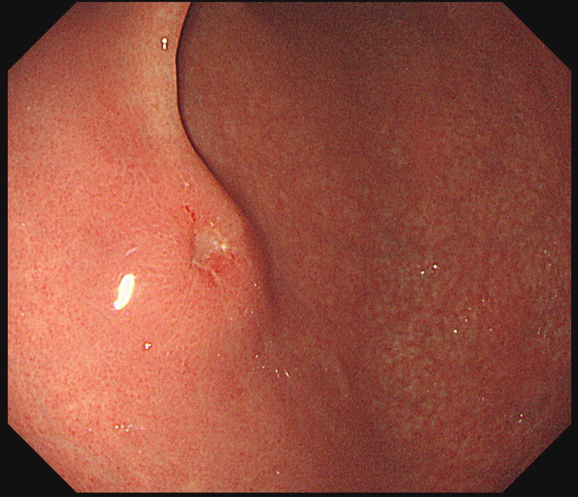
In the screening endoscopy for a young adult, a small elevated with central depression was found. The edge was discrete except hyperemia at 3 to 6 o'clock direction. The pathology was poorly cohesive carcinoma with signet ring cell feature.
Repeated endoscopy after referral showed similar images. The endoscopist (1st year fellow) guessed it would be an AGC, but I was not sure.
Advanced gastric cancer B-I
#1 6(AW of low body)
- Location : anterior wall of low body
- Size : 2 cm
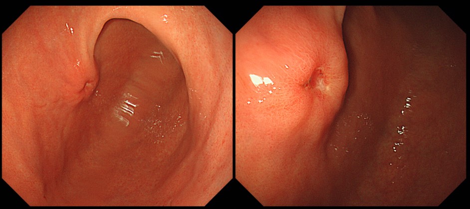
Final patholgy after surgery was as follows.
Stomach, subtotal gastrectomy: Advanced gastric carcinoma
1. Location : lower third, Center at body and anterior wall
2. Gross type : Borrmann type 2
3. Histologic type : tubular adenocarcinoma, poorly (poorly cohesive) differentiated
4. Histologic type by Lauren : diffuse
5. Size : 1.8x1.5 cm
6. Depth of invasion : invades muscularis propria (pT2)
7. Resection margin: free from carcinoma, safety margin: proximal 3.4 cm, distal 5 cm
8. Lymph node metastasis : metastasis to one out of 47 regional lymph nodes (pN1), (perinodal extension: absent) (1/47: "3", 1/11; "4", 0/11; "5", 0/2; "6", 0/5; "7", 0/4; "9", 0/2; "8a", 0/4; "11p", 0/5; "12a", 0/1; "4sb", 0/1; "1", 0/1)
9. Lymphatic invasion : not identified
10. Venous invasion : not identified
11. Perineural invasion : not identified
12. Peritoneal cytology : negative
You did a good job, Dr. Yoo.
© 일원내시경교실 바른내시경연구소 이준행. EndoTODAY Endoscopy Learning Center. Lee Jun Haeng. (2019-9-14)