EndoTODAY 내시경 교실
Beginner | ESA | Schedule | OPD
Seminars | Atlas | Recent | Links
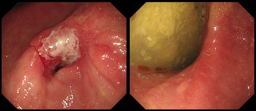
 [Gastric cancer 835. Stricture after ESD]
001 | 101 | 201 | 301 | 401 | 501 | 601 | 701 | 801 | 901 | 1000
An ill-defined mucosal lesion near the pylorus was found in the screening endoscopy of a middle-aged woman. ESD was done.
(2018)
ESD : Early gastric carcinoma
1. Location : antrum, posterior wall
2. Gross type : EGC type IIb
3. Histologic type : tubular adenocarcinoma, moderately differentiated
4. Histologic type by Lauren : intestinal
5. Size of carcinoma : (1) longest diameter, 60 mm (2) vertical diameter, 32 mm
6. Depth of invasion : invades submucosa, (depth of sm invasion : 500 ㎛) (pT1b)
7. Resection margin : free from carcinoma(N)
8. Lymphatic invasion : not identified(N)
9. Venous invasion : not identified(N)
10. Perineural invasion : not identified(N)
11. Microscopic ulcer : absent
12. Histologic heterogeneity: absent
In the follow-up endoscopy, stricture was found.
I made a report as followings and notice to the duty doctor.
"오전에 음식물이 남아있어서 늦은 오후에 다시 시술한 것입니다. 위체부의 다량의 음식 물이 관찰됩니다. Left decubitus로 검사하였으므로 전정부는 관찰할 수 있습니다. ESD ulcer 가 아직 관찰되고 이로 인하여 현저히 좁아져 3-4mm 정도 밖에 lumen이 보이지 않을 정도로 좁아졌습니다. 재발 소견은 없으나 조직검사 2개 하였습니다. 헬리코박터 검사는 할 수 없었습니다. (음식물 때문)"
© 일원내시경교실 바른내시경연구소 이준행. EndoTODAY Endoscopy Learning Center. Lee Jun Haeng. (2020-3-2)