 EndoTODAY 내시경 교실
Beginner | ESA | Schedule | OPD
Seminars | Atlas | Recent | Links
 [Gastric cancer 768. EGC]
001 | 101 | 201 | 301 | 401 | 501 | 601 | 701 | 801 | 901 | 1000
2017년 환자입니다. 80대 중반 남성입니다. 10년 전 관상동맥 stent 후 약을 드시고 계신 분이셨습니다.
개업가에서 내시경 검사 후 조직검사에서 adenoma가 나와 6개월 후 내시경 재검을 권유받았는데 큰 병원에서 2차 의견을 듣고 싶다고 오셨습니다. Fujifilm 내시경 사진이었는데 다소 희미하였지만 약간의 함몰부가 있고 전벽쪽으로는 융기된 부위도 보였습니다. 일단 외부슬라이드 재판독을 하였는데 well-differentiated adenocarcinoma로 보고되어 즉시 ESD를 시행하였습니다.
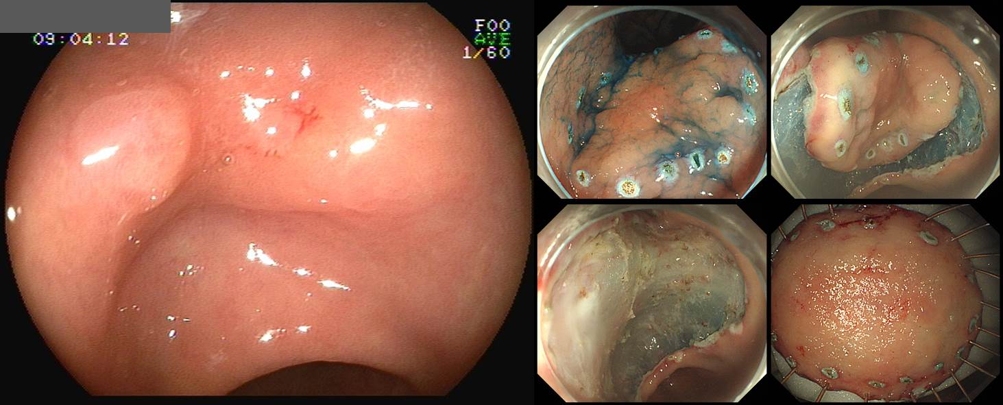
ESD: Early gastric carcinoma
1. Location : proximal antrum, lesser curvature
2. Gross type : EGC type IIa+IIb
3. Histologic type : tubular adenocarcinoma, well differentiated
4. Histologic type by Lauren : intestinal
5. Size of carcinoma : (1) longest diameter, 44 mm (2) vertical diameter, 23 mm
6. Depth of invasion : invades mucosa (muscularis mucosa) (pT1a)
7. Resection margin : free from carcinoma(N), safety margin : distal 2 mm, proximal 5 mm, anterior 4 mm, posterior 8 mm, deep 200 ㎛
8. Lymphatic invasion : not identified(N)
9. Venous invasion : not identified(N)
10. Perineural invasion : not identified(N)
11. Microscopic ulcer : absent
12. Histologic heterogeneity: absent
13. Associated finding: Gastritis cystica superficialis
장경 44mm로 제법 큰 조기위암이었지만 침윤 깊이는 점막암이었습니다. 수술 없이 추적 관찰을 하고 있습니다.
관상동맥 stent 병력이 있는 80대 중반 남성에서 건강검진 내시경이 필요한지 명확하지 않지만 일단 발견된 위암은 치료하지 않을 수 없다고 생각합니다.
내시경에서 focal lesion이 발견되면 주변과의 경계를 평가하여 크기를 짐작하는 습관을 가지는 것이 좋겠습니다. 비록 조직검사에서 선종으로 나왔더라도 크기가 4cm면 쉽게 follow up을 권하지는 않았을 것 같습니다.
© 일원내시경교실 바른내시경연구소 이준행. EndoTODAY Endoscopy Learning Center. Lee Jun Haeng. (2019-7-20)