EndoTODAY 내시경 교실
Beginner | ESA | Schedule | OPD
Seminars | Atlas | Recent | Links
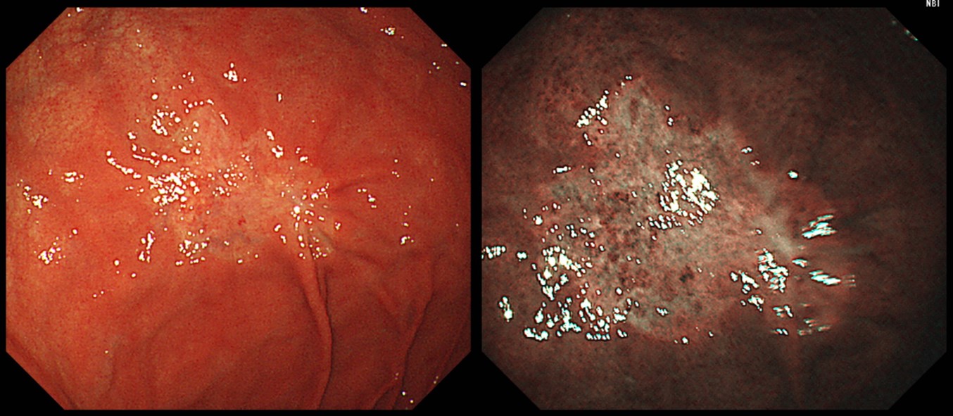
![]() [Gastric cancer 808. EGC (fundus)]
[Gastric cancer 808. EGC (fundus)]
001 | 101 | 201 | 301 | 401 | 501 | 601 | 701 | 801 | 901 | 1000
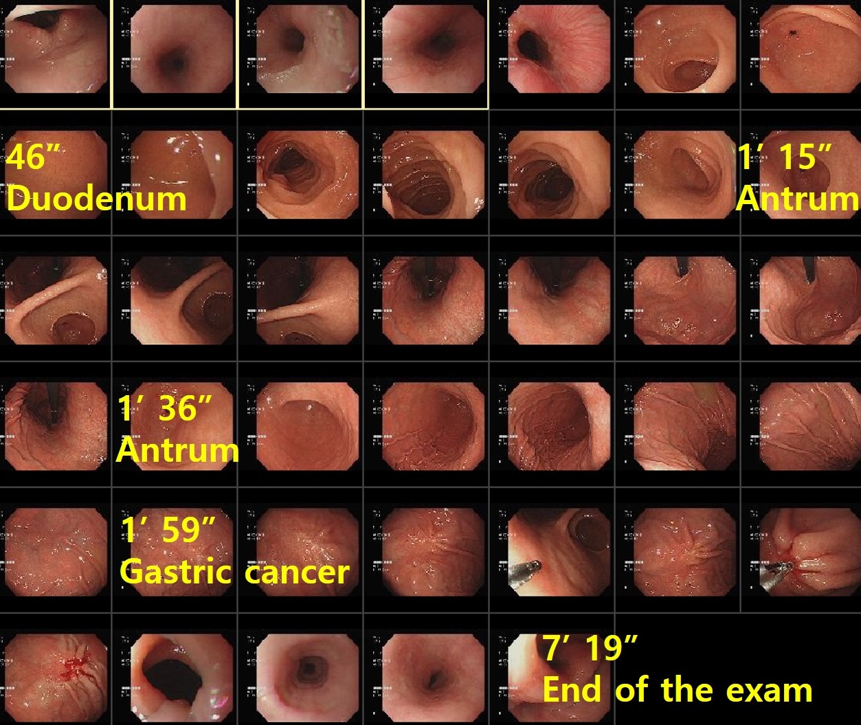
A slightly depressed discolorated EGC was found during a screening endoscopy. The initial biopsy was "atypical signet ring cells." After referral, the repeated biopsy was poorly differentiated adenocarcinoma, and radical total gastrectomy was done.
Stomach, radical total gastrectomy: early gastric carcinoma
1. Location : upper third, Center at fundus and posterior wall
2. Gross type : EGC type IIc
3. Histologic type : tubular adenocarcinoma, poorly (solid) differentiated
4. Histologic type by Lauren : diffuse
5. Size : 1.5x1.5 cm
6. Depth of invasion : invades mucosa (muscularis mucosae) (pT1a)
7. Resection margin: free from carcinoma, safety margin: proximal 5.0 cm, distal 20.1 cm
8. Lymph node metastasis : no metastasis in 56 regional lymph nodes (pN0)
9. Lymphatic invasion : not identified
10. Venous invasion : not identified
11. Perineural invasion : not identified
12. Peritoneal cytology : negative
13. AJCC stage by 8th edition: pT1a N0
When I reviewed the endoscopic images of the screening endoscopy, I found that the endoscopist has a very good routine pattern of examinations.
Thank you very much for your great job. You really did a lot to the patient.
© 일원내시경교실 바른내시경연구소 이준행. EndoTODAY Endoscopy Learning Center. Lee Jun Haeng. (2019-11-14)