 EndoTODAY 내시경 교실
Beginner | ESA | Schedule | OPD
Seminars | Atlas | Recent | Links
![]() [Gastric cancer 807. EGC (fundic gland type)]
[Gastric cancer 807. EGC (fundic gland type)]
001 | 101 | 201 | 301 | 401 | 501 | 601 | 701 | 801 | 901 | 1000
Follow up endoscopy for a 50 years old male with a polyp (previous biopsy: hyperplastic polyp) was done and the forceps biopsy was atypical proliferation of fundic glands, suggestive of tubular adenocaricnoma, well differentiated, fundic gland type. ESD was done under the impression of r/o EGC.
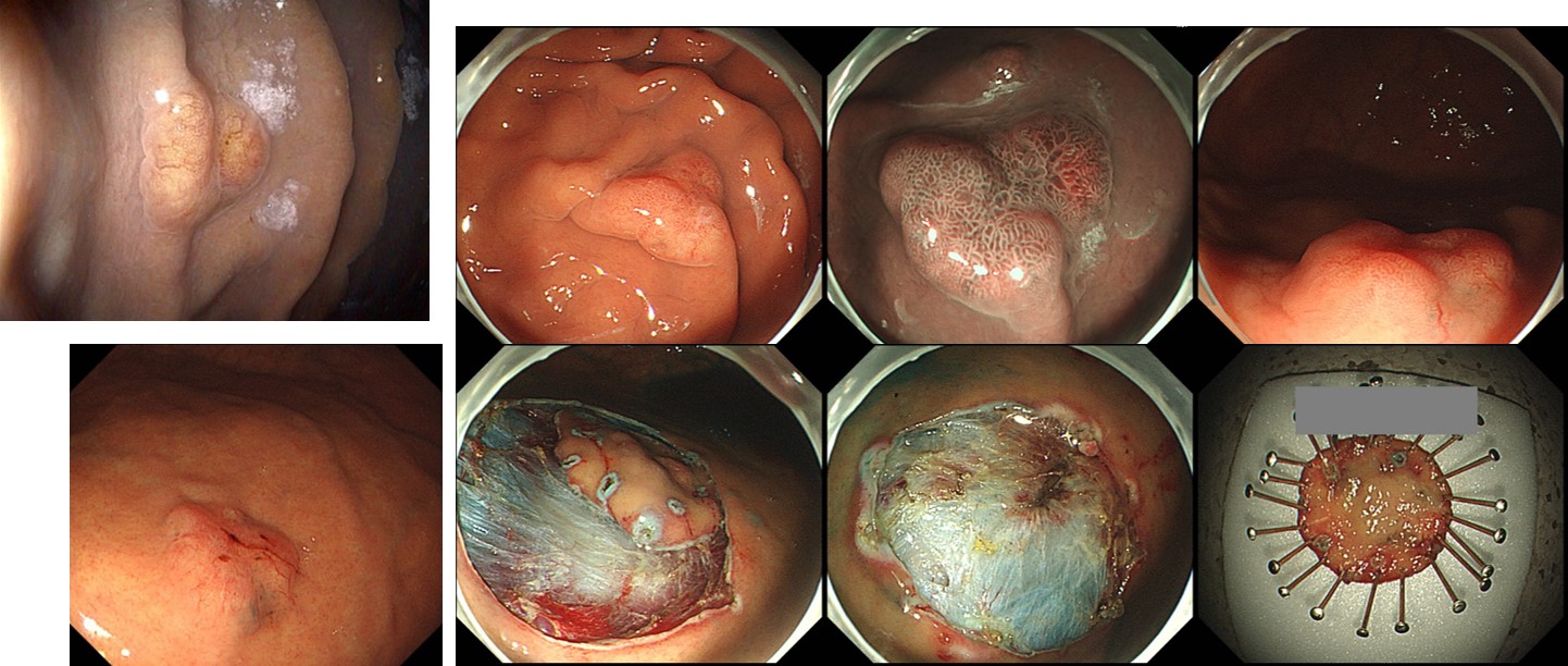
ESD: Early gastric carcinoma
1. Location : high body, greater curvature
2. Gross type : EGC type IIa
3. Histologic type : tubular adenocarcinoma, W/D (fundic gland type)
4. Histologic type by Lauren : intestinal
5. Size of carcinoma : (1) longest diameter, 18 mm (2) vertical diameter, 11 mm
6. Depth of invasion : invades submucosa, (depth of sm invasion : 300 ) (pT1b)
7. Resection margin : free from carcinoma(N) safety margin : distal 5 mm, proximal 5 mm, anterior 12 mm, posterior 1 mm, deep 50
8. Lymphatic invasion : not identified(N)
9. Venous invasion : not identified(N)
10. Perineural invasion : not identified(N)
11. Microscopic ulcer : absent
12. Histologic heterogeneity: absent
I suspect it may be a case of gastric cancer arising from a fundic gland polyp.
© 일원내시경교실 바른내시경연구소 이준행. EndoTODAY Endoscopy Learning Center. Lee Jun Haeng. (2019-11-8)