EndoTODAY 내시경 교실
Beginner | ESA | Schedule | OPD
Seminars | Atlas | Recent | Links
![]() [Gastric cancer 833. Tumor island]
[Gastric cancer 833. Tumor island]
001 | 101 | 201 | 301 | 401 | 501 | 601 | 701 | 801 | 901 | 1000
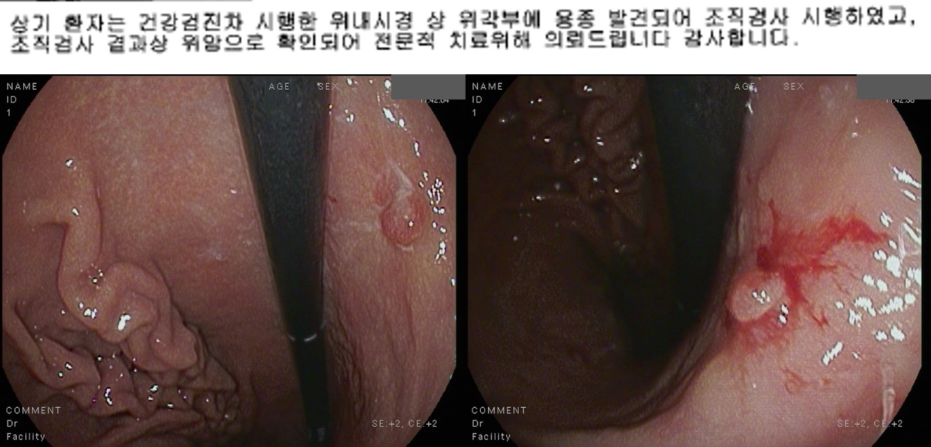
A patient with a gastric polyp, which was found in the screening endoscopy, was referred for the endoscopic treatment.
When I reviewed the outside endoscopic images, it was not a polypoid EGC, but a depressed type EGC with a small elevated lesion (tumor island). Endoscopic resection was done as usual.

ESD: Early gastric carcinoma
1. Location : angle, posterior wall
2. Gross type : EGC type IIc
3. Histologic type : tubular adenocarcinoma, moderately differentiated
4. Histologic type by Lauren : intestinal
5. Size of carcinoma : (1) longest diameter, 18 mm (2) vertical diameter, 14 mm
6. Depth of invasion : invades mucosa (lamina propria) (pT1a)
7. Resection margin : free from carcinoma(N), safety margin : distal 11 mm, proximal 4 mm, anterior 12 mm, posterior 6 mm, deep 300 ㎛
8. Lymphatic invasion : not identified(N)
9. Venous invasion : not identified(N)
10. Perineural invasion : not identified(N)
11. Pre-existing adenoma : none
12. Microscopic ulcer : absent
13. Histologic heterogeneity: absent
It is easier to find elevated lesions than depressed lesions. When a lesion was found, we need to see the surrounding mucosa carefully.
You can see more images at EndoTODAY tumor island.
© 일원내시경교실 바른내시경연구소 이준행. EndoTODAY Endoscopy Learning Center. Lee Jun Haeng. (2020-2-20)