EndoTODAY 내시경 교실
Beginner | ESA | Schedule | OPD
Seminars | Atlas | Recent | Links
![]() [Gastric cancer 861. ESD for EGC (46mm)]
[Gastric cancer 861. ESD for EGC (46mm)]
001 | 101 | 201 | 301 | 401 | 501 | 601 | 701 | 801 | 901 | 1000
A lady was referred for an endoscopic treatment of a small early gastric cancer. In the ESD procedure, I found that the lesion was larger than expected. I examined the margin of the lesion very carefully with i-Scan mode of Pentax Imagina endoscope and indigocarmine chromoendoscopy. ESD procedure was done as usual.
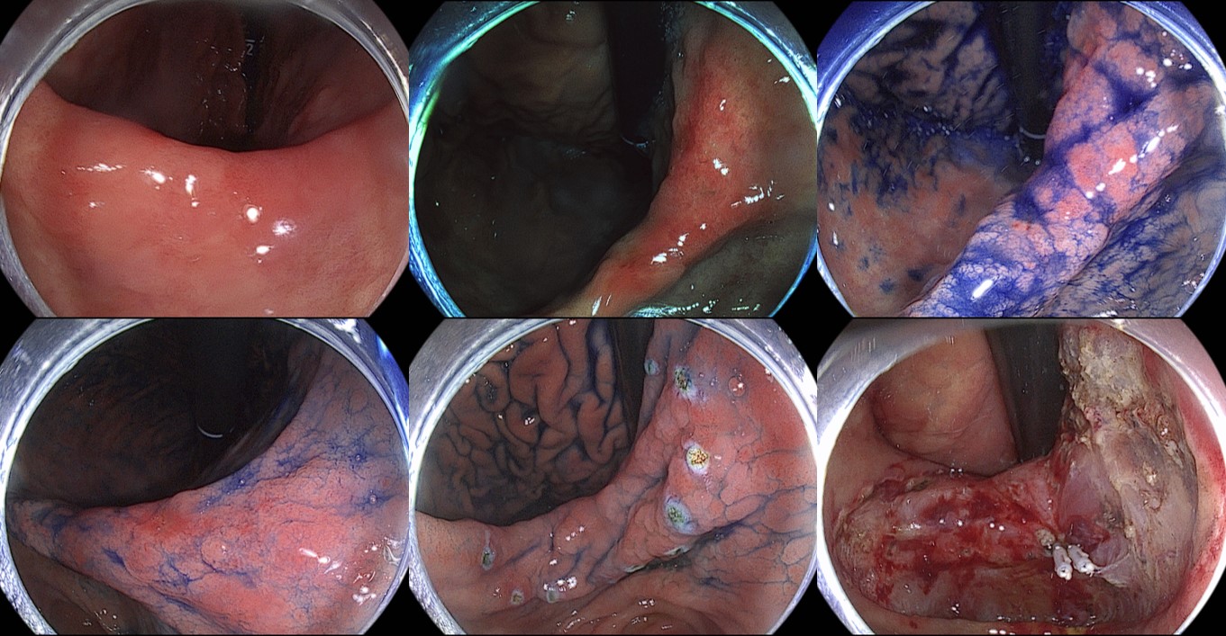
ESD: Early gastric carcinoma
1. Location : angle, lesser curvature
2. Gross type : EGC type IIb + IIc
3. Histologic type : tubular adenocarcinoma, moderately differentiated (WHYX type)
4. Histologic type by Lauren : intestinal
5. Size of carcinoma : (1) longest diameter, 46 mm (2) vertical diameter, 30 mm
6. Depth of invasion : invades mucosa (muscularis mucosa) (pT1a)
7. Resection margin : free from carcinoma(N), safety margin : distal 10 mm, proximal 2 mm, anterior 12 mm, posterior 4 mm, deep 300 ㎛
8. Lymphatic invasion : not identified(N)
9. Venous invasion : not identified(N)
10. Perineural invasion : not identified(N)
11. Microscopic ulcer : absent
12. Histologic heterogeneity: absent
You can see the marking step in the following video clip.
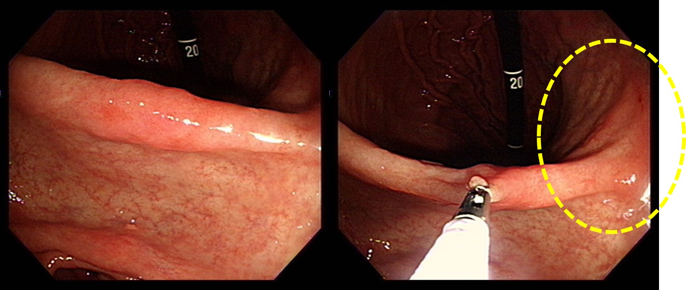
In the review of the first endoscopy images, an area of discoloration was suspected in the posterior aspect of the angle (yellow ring).
내시경 검사에서 가장 중요한 것은 존재진단입니다. 암을 찾는 것이 가장 중요합니다.
그러나 존재진단은 출발에 지나지 않습니다. 좋은 치료를 위해서는 성격 진단도 빠질 수 없습니다. 병소의 크기와 모양을 정확히 평가하는 것이 중요한 이유입니다. Focal lesion이 발견되면 크기가 얼마일지 평가하는 것은 아무리 강조해도 지나치지 않습니다.
© 일원내시경교실 바른내시경연구소 이준행. EndoTODAY Endoscopy Learning Center. Lee Jun Haeng. (2020-5-30)