EndoTODAY 내시경 교실
Beginner | ESA | Schedule | OPD
Seminars | Atlas | Recent | Links
 [Gastric cancer 650 - Residual cancer in surgery for noncurated ESD]
001 | 101 | 201 | 301 | 401 | 501 | 601 | 701 | 801 | 901 | 1000
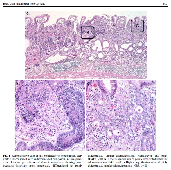
The rate of pathological non-curative resection of EGC by ESD is about 10-20%. In additional surgery for non-curative resection cases, local residual tumor is found in 5-10%, and lymph node metastasis is also found in 5-10%. In this case of histological heterogeneity, lateral margin was involved. Surgery was done and 1.2cm x 0.3cm sized residual tumor in lamina propria was found.
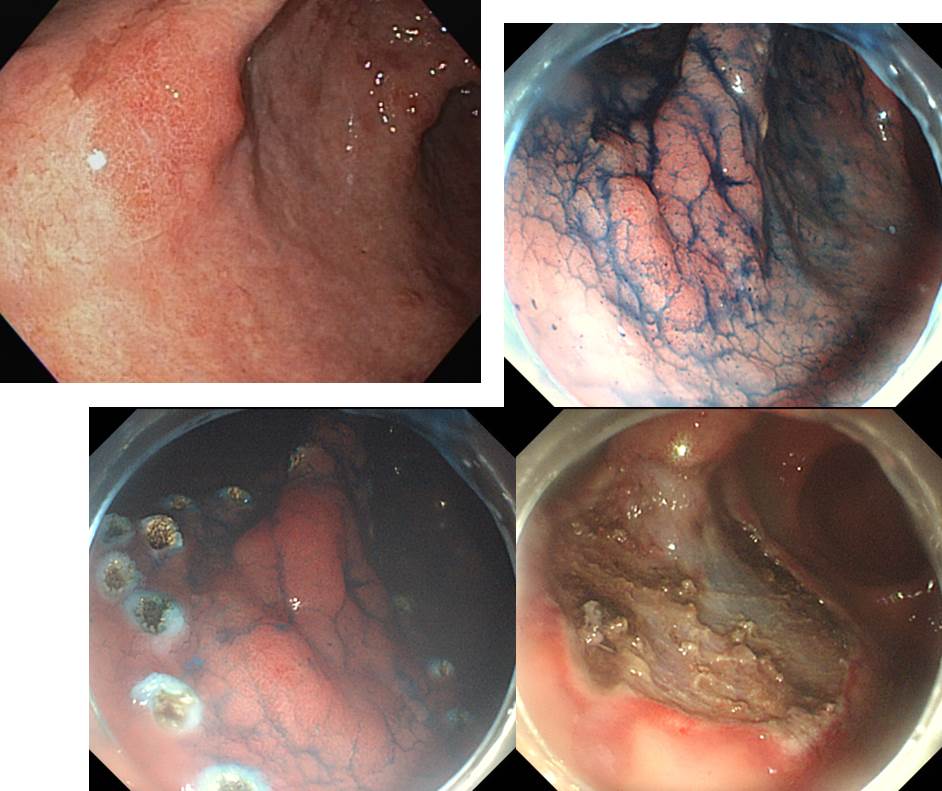
ESD: Early gastric carcinoma
1. Location : angle, anterior wall
2. Gross type : EGC type IIa+IIc
3. Histologic type : tubular adenocarcinoma, well differentiated (WHYX type) >> tubular adenocarcinoma, poorly differentiated (10%) > signet ring cell carcinoma (10%)
4. Histologic type by Lauren : mixed
5. Size of carcinoma : (1) longest diameter, 28 mm (2) vertical diameter, 26 mm
6. Depth of invasion : invades mucosa (lamina propria) (pT1a)
7. Resection margin : involvement of proximal margin by carcinoma (poorly differentiated) safety margin : distal 6 mm, proximal 0 mm, anterior 8 mm, posterior 6 mm, deep 1000 ㎛
8. Lymphatic invasion : not identified(N)
9. Venous invasion : not identified(N)
10. Perineural invasion : not identified(N)
11. Microscopic ulcer : absent
12. Histologic heterogeneity: present
Resectio margin positive에서 ESD나 ablation을 추가하기도 하고 수술을 권할 수도 있습니다. 경계가 불분명한 부위였고, resection margin 부위가 poorly differentiated component였으므로 수술을 권했습니다.
Stomach, subtotal gastrectomy: Status post ESD, Early gastric carcinoma, residual;
1. Location : lower third, Center at angle and anterior wall
2. Gross type : EGC type (unspecified)
3. Histologic type : tubular adenocarcinoma, well differentiated
4. Histologic type by Lauren : intestinal
5. Size : 1.2x0.3 cm
6. Depth of invasion : invades mucosa (lamina propria) (pT1a)
7. Resection margin: free from carcinoma, safety margin: proximal 4.0 cm, distal 7.2 cm
8. Lymph node metastasis : no metastasis in 21 regional lymph nodes (pN0)
9. Lymphatic invasion : not identified
10. Venous invasion : not identified
11. Perineural invasion : not identified
12. AJCC stage by 8th edition (ESD+resection) : pT1a N0
[2018-7-28. 애독자 질문]
미분화 혼재암 ESD 후 절제변연 양성으로 수술을 권한 증례(EndoTODAY 위암 650)에 대한 질문입니다. Mixed type 의 경우 악성의 가능성이 더 높다고 하신 바 있습니다 (Shim CN. Surg Endosc 2015). 일전에 보여주신 환자설명자료(EndoTODAY 위암 615)에서는 '최근에는 혼재형에서도 ESD를 할 수 있다'고 하셨기에 조금 혼란스럽습니다.
[2018-7-28. 이준행 답변]
위암을 조직형에 따라 분화형과 미분화형으로 나누는 것은 엄밀한 방법은 아니지만, 편리한 측면이 있어서 ESD 영역에서 종종 사용되고 있습니다. 분화형과 미분화형이 섞인 미분화 혼재암은 아직 그 정의조차 명확하지 않습니다. Histological heterogeneity라고 표현되기도 합니다. 같은 말입니다.
미분화 혼재암이 분화조직형보다 나쁜 것은 틀림없습니다. 그런데, 미분화 혼재암을 순수한 미분화 조직형 암과 비교하면 어떨까요? 직관적으로는 미분화 혼재형은 분화조직형과 미분화조직형의 중간 정도일 것 같지만, 연구 결과는 섞인 것은 순순한 것보다 나쁘다는 것입니다 (Shim CN. Surg Endosc 2015). 저도 미분화 혼재암은 순수한 미분화 조직형 (P/D 혹은 SRC) 위암보다 비슷하거나 더 나쁠 것으로 생각하고 있습니다.
내시경 육안 소견이 분화조직형 위암에 가까운데 조직검사에서 mixed type으로 나오면 조심스럽게 ESD를 시행할 수 있습니다. 물론 크기 기준을 엄격하게 적용합니다. 1-2cm 이상에서는 절대 시행하지 않는 것이지요.
임상에서는 ESD 전 조직검사에서는 분화조직형 위암으로 나왔는데 ESD 후 최종 병리결과가 미분화 혼재형으로 나온 경우가 많습니다.
(1) 미분화 혼재암이더라도 점막암이고 절제 변연이 음성이면 완전절제로 간주하고 경과관찰을 합니다.
(2) 미분화 혼재암이고 점막하암이면 수술하는 경우가 많습니다. 이 부분은 다소 논란이 있어 case by case로 접근합니다. 일반적인 curative resection criteria에 만족하면 경과관찰을 하기도 합니다.
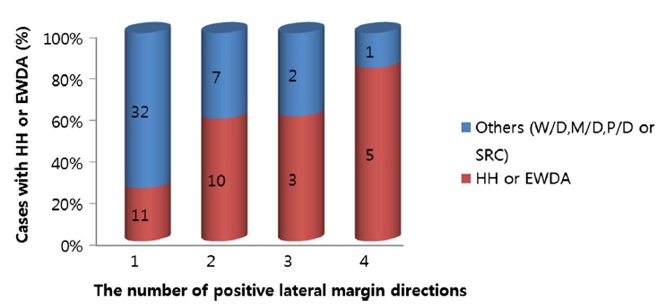
(3) 문제는 미분화 혼재암이고 절제 변연이 양성인 경우입니다. 분화조직형 위암에 절제변연이 양성이면 추가 절제술을 하거나, 소작술을 하거나, 조심스럽게 경과관찰을 할 수 있습니다. 미분화 혼재암에서 절제 변연이 양성이면 residual tumor의 가능성이 높습니다. 미분화 혼재암은 내시경 육안소견으로 위암의 경계를 명확히 확인하기 어렵고, ESD 후 multiple lateral margin involvement가 자주 발견됩니다 (Lee JH. Surg Endosc 2015). 저는 미분화 혼재암인데 절제변연이 양성이면, 특히 미분화암이 절제변연에서 발견되면 수술을 권하고 있습니다. 문의하신 EndoTODA 위암 650의 병리결과를 보면 "involvement of proximal margin by carcinoma (poorly differentiated)"라는 부분이 있었습니다. 수술 후 최종 병리에서도 residual tumor가 있는 것으로 나와서 수술을 권하길 잘 했다고 생각하였습니다.
저와 함께 일하고 있는 민병훈 교수께서 삼성서울병원의 미분화 혼재암 ESD 성적에 대하여 보고한 바 있으니 참고하시기 바랍니다 (Min BH. Gastric Cancer 2015).
Min. 2015
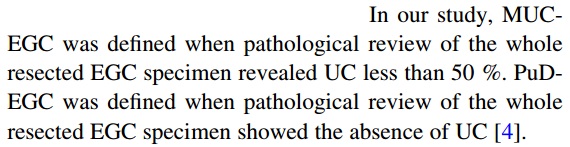
En bloc resection and en bloc with R0 resection rates in MUC-EGC cases were 94.1 % and 81.7 %, respectively. MUC-EGC was significantly associated with larger tumor size, more frequent submucosal invasion, and lymphovascular invasion compared to PuD-EGC. Despite these aggressive features of MUC-EGC, no lymph node metastasis or extragastric recurrence occurred during follow-up after ESD if MUC-EGC met the curative endoscopic resection (ER) criteria for tumors of absolute or expanded indications.
 [References]
© 일원내시경교실 바른내시경연구소 이준행. EndoTODAY Endoscopy Learning Center. Lee Jun Haeng. (2018-7-26)